- Главная
- Разное
- Дизайн
- Бизнес и предпринимательство
- Аналитика
- Образование
- Развлечения
- Красота и здоровье
- Финансы
- Государство
- Путешествия
- Спорт
- Недвижимость
- Армия
- Графика
- Культурология
- Еда и кулинария
- Лингвистика
- Английский язык
- Астрономия
- Алгебра
- Биология
- География
- Детские презентации
- Информатика
- История
- Литература
- Маркетинг
- Математика
- Медицина
- Менеджмент
- Музыка
- МХК
- Немецкий язык
- ОБЖ
- Обществознание
- Окружающий мир
- Педагогика
- Русский язык
- Технология
- Физика
- Философия
- Химия
- Шаблоны, картинки для презентаций
- Экология
- Экономика
- Юриспруденция
Immunology of Dermatophytes and Dermatophytosis презентация
Содержание
- 1. Immunology of Dermatophytes and Dermatophytosis
- 2. Agent Properties Dermatophytes are complex fungi
- 3. Aetiology Over 90% of feline
- 4. Epidemiology Arthrospores are transmitted through contact
- 5. Pathogenesis Healthy skin acts as an effective
- 6. The incubation period of ringworm caused
- 7. Predisposing factors Young age (first 2
- 8. In general, the zoophilic species cause
- 9. Immunity Ringworm rarely recurs, suggesting an
- 10. Although dermatophyte infection is confined to
- 11. Antibodies In spite of the superficial nature
- 12. Citron was the first to report
- 13. Several investigators have demonstrated the production
- 14. Investigations showed that inoculation of living
- 15. Nonspecific Resistance Many nonspecific factors may account
- 16. The dermatophytes do not usually grow
- 17. Therapy and disease management In immunocompetent
- 18. Topical treatment is less effective in
- 19. Systemic therapy Griseofulvin In some countries, the
- 20. Вакцина содержит антиген из штамма Microsporum
- 22. www.themegallery.com Thank You!
Слайд 2Agent Properties
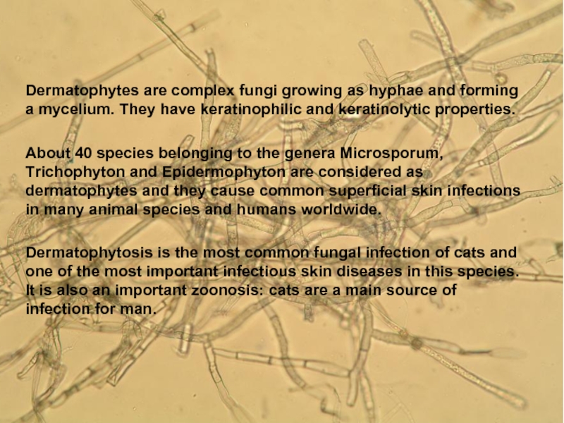
Dermatophytes are complex fungi growing as hyphae and forming
a mycelium. They have keratinophilic and keratinolytic properties.
About 40 species belonging to the genera Microsporum, Trichophyton and Epidermophyton are considered as dermatophytes and they cause common superficial skin infections in many animal species and humans worldwide.
Dermatophytosis is the most common fungal infection of cats and one of the most important infectious skin diseases in this species. It is also an important zoonosis: cats are a main source of infection for man.
Слайд 3Aetiology
Over 90% of feline dermatophytosis cases worldwide are caused by
Microsporum canis .
Others are caused by infection with M. gypseum, Trichophyton mentagrophytes, T. quinckeanum, T. verrucosum or other agents. With the exception of M. gypseum, all produce proteolytic and keratolytic enzymes that enable them to utilize keratin as the sole source of nutrition after colonization of the dead, keratinized portion of epidermal tissue (mostly stratum corneum and hairs, sometimes nails).
By segmentation and fragmentation of the hyphae, dermatophytes produce arthrospores, which are highly resistant, surviving in a dry environment for 12 months or longer . In a humid environment, however, arthrospores are short-lived. High temperatures (100°C) destroy them quickly.
Слайд 4Epidemiology
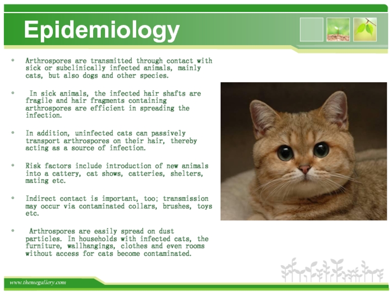
Arthrospores are transmitted through contact with sick or subclinically infected
animals, mainly cats, but also dogs and other species.
In sick animals, the infected hair shafts are fragile and hair fragments containing arthrospores are efficient in spreading the infection.
In addition, uninfected cats can passively transport arthrospores on their hair, thereby acting as a source of infection.
Risk factors include introduction of new animals into a cattery, cat shows, catteries, shelters, mating etc.
Indirect contact is important, too; transmission may occur via contaminated collars, brushes, toys etc.
Arthrospores are easily spread on dust particles. In households with infected cats, the furniture, wallhangings, clothes and even rooms without access for cats become contaminated.
Слайд 5Pathogenesis
Healthy skin acts as an effective physical barrier against fungal invasion.
The increased rate of regeneration of epidermal cells in response to contact with the dermatophyte with the consequent removal of fungus from the skin surface is another protective mechanism. As dermatophytes cannot penetrate healthy skin, many cats are merely passive carriers of the arthrospores or remain subclinically infected. Whether such an infection will lead to clinical signs depends on endogenous and exogenous factors.
Слайд 6
The incubation period of ringworm caused by M. canis is 1
to 3 weeks. During this time, hyphae grow along the hair shafts through the stratum corneum to the follicles where they produce spores that form a thick layer around the hair shafts.
As dermatophytes are susceptible to high temperatures, they cannot colonize deeper parts of the skin or the follicle itself.
Therefore, the hair grows normally but breaks easily near the skin surface resulting in hair loss.
Several metabolic products of the fungus may induce an inflammatory skin response and may be observed mainly around the infected area, forming sometimes ring-like lesions with central areas of healing and papules on the periphery (“ringworm”).
On rare occasions, an inflammatory reaction to hyphae induces a nodular granulomatous lesion involving dermis and draining on the skin surface. These so-called pseudomycetomas are more often seen in Persian cats, even concurrently with classical lesions.
Слайд 7Predisposing factors
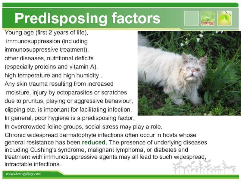
Young age (first 2 years of life),
immunosuppression (including
immunosuppressive treatment),
other diseases, nutritional deficits
(especially proteins and vitamin A),
high temperature and high humidity .
Any skin trauma resulting from increased
moisture, injury by ectoparasites or scratches
due to pruritus, playing or aggressive behaviour,
clipping etc. is important for facilitating infection.
In general, poor hygiene is a predisposing factor.
In overcrowded feline groups, social stress may play a role.
Chronic widespread dermatophyte infections often occur in hosts whose general resistance has been reduced. The presence of underlying diseases including Cushing's syndrome, malignant lymphoma, or diabetes and treatment with immunosuppressive agents may all lead to such widespread, intractable infections.
Слайд 8
In general, the zoophilic species cause more inflammatory infections which may
heal spontaneously and result in relative resistance to reinfection.
The anthropophilic species usually cause more chronic, less circumscribed infections which result in less resistance to reinfection.
The anthropophilic species usually cause more chronic, less circumscribed infections which result in less resistance to reinfection.
Слайд 9Immunity
Ringworm rarely recurs, suggesting an effective and long-lasting immunity. Experimental
studies confirm that animals express increased resistance to subsequent challenge by the homologous fungus. Re-infections may occur, but require a much greater number of spores, and these subsequent infections are usually cleared more rapidly . It has been suggested that for the development of full immunity, the infection must run its natural course, as in cats whose infection was aborted with antifungal treatment, the delayed type hypersensitivity reactions were often weaker.
Слайд 10
Although dermatophyte infection is confined to the superficial keratinised tissues, humoral
and cellular immune responses are induced.
Prominent activation of T helper type 2 cells and the corresponding cytokine profile leads to antibody formation followed by chronic disease, whereas activation of Th1 cells stimulates a cell-mediated response characterized by the cytokines interferon-γ (IFN-γ), interleukin 12, and IL-2, and leads to recovery. This implies that cats are protected against reinfection.
The role of the humoral response in dermatophytosis is unclear, although specific antibodies could have a direct fungistatic effect by means of opsonization and complement activation .
Слайд 11Antibodies
In spite of the superficial nature of most infections, circulating antibodies
have been demonstrated in sera from both animals and humans with either natural or experimental dermatophytosis.
Disagreement has arisen among some investigators as to their presence or the specificity of these antibodies for the infecting organism. The differences in experimental results may be accounted for by
the variety of methods used for analyses, including Prausnitz-Kiistner, agglutination, precipitation, complement-fixation, immunofluorescence, and "in vitro" growth inhibition tests;
the different test antigens used, including concentrated culture filtrates, commercial "trichophytins," mycelial extracts and crude polysaccharides, or other fractions;
the time during the course of infection at which blood was drawn from the host; and
the nature of the infection (chronic versus acute).
Слайд 12
Citron was the first to report the presence of circulating antibodies
to dermatophytes in sera of experimental animals. He injected rabbits with suspensions of live T. gypseum and detected precipitating antibodies to extracts prepared from this species. The antibodies also reacted with extracts of Achorion schoenleinii and A. quinckeanum. His studies were followed by several unsuccessful attempts to demonstrate antibodies in sera of animals with dermatophytosis . Verotti was the first to demonstrate circulating antibodies in serum from an animal with a natural cutaneous infection. He detected complement-fixing antibodies to M. lanosum in the serum of a dog. These antibodies also reacted with other species of dermatophytes.
Слайд 13
Several investigators have demonstrated the production of agglutinins, precipitins, and complement-fixing
antibodies by immunization of animals, especially rabbits, with killed dermatophyte cells.
The earliest reports were those of Kolmer and Strickler , Fuke , and Sharp .
Precipitins were frequently detected, appearing after 24 days following infection and persisting up to 13 weeks. Low complement-fixing antibody titers rose after several reinfections.
The earliest reports were those of Kolmer and Strickler , Fuke , and Sharp .
Precipitins were frequently detected, appearing after 24 days following infection and persisting up to 13 weeks. Low complement-fixing antibody titers rose after several reinfections.
Слайд 14
Investigations showed that inoculation of living or killed dermatophyte cells by
routes other than the cutaneous could produce hypersensitivity and resistance to subsequent cutaneous infection. However, injection of guinea pigs with whole cells by intracardial, intramuscular, subcutaneous, or intraperitoneal routes induced only a poor or partial protection. Subsequent cutaneous infections took an abortive course, but protection was of relatively short duration. No protection was obtained by injection of culture filtrates . Hypersensitivity and partial resistance could also be obtained in guinea pigs by rubbing their skin with killed mycelium or by repeated intradermal injections of a "toxic substance" obtained from the supernatant fluid of mixtures of extracts of infected skin and mycelium incubated at 37 C for 24 h .
Later, Wharton et al. injected rabbits subcutaneously with a killed T. rubrum suspension in Freund complete adjuvant. The immunized rabbits were completely resistant to infection for up to 17 months or more. This resistance was greater than that obtained in rabbits after cutaneous infections. Precipitating antibodies were detected in the resistant animals. Injection of culture filtrate or extract of the fungus did not give protection.
Later, Wharton et al. injected rabbits subcutaneously with a killed T. rubrum suspension in Freund complete adjuvant. The immunized rabbits were completely resistant to infection for up to 17 months or more. This resistance was greater than that obtained in rabbits after cutaneous infections. Precipitating antibodies were detected in the resistant animals. Injection of culture filtrate or extract of the fungus did not give protection.
Слайд 15Nonspecific Resistance
Many nonspecific factors may account for natural resistance to infection
in humans or animals. This slide is limited mainly to the "serum factor," a fungistatic substance in serum of normal individuals and animals.
This factor is believed to limit the growth of the dermatophytes to the keratinized layers, i.e., prevent their invasion of living tissues. Several investigators have demonstrated the antidermatophytic activity of normal serum. Sera of newborns, adults, and animals show this inhibitory activity.
This factor is believed to limit the growth of the dermatophytes to the keratinized layers, i.e., prevent their invasion of living tissues. Several investigators have demonstrated the antidermatophytic activity of normal serum. Sera of newborns, adults, and animals show this inhibitory activity.
Слайд 16
The dermatophytes do not usually grow in internal organs of the
living body. However, they can easily be cultured on these organs once they have been removed.
Blank et al. cultured human skin, both newborn and adult, as an organized tissue and infected the skin with dermatophytes to simulate natural human infection. After incubation with or without serum, the infected tissues were sectioned and the localization of the dermatophytes in the tissues was studied.
The authors found that the hyphae readily invaded all layers of the skin when no serum was present, but that the growth of the fungus was restricted to the keratinized layers of the skin when serum had been added to the culture medium. This activity was lost after dialysis or heating of the serum at 56 C.
The "serum factors" were not identified, but neither gamma globulin nor other protein fractions, such as albumin and Cohn fractions II, III, or IV, showed any activity.
The inhibitor from human serum was isolated and identified as alpha-2 macroglobulin. Its significance in the process of dermatophytosis is not yet known.
Слайд 17Therapy and disease management
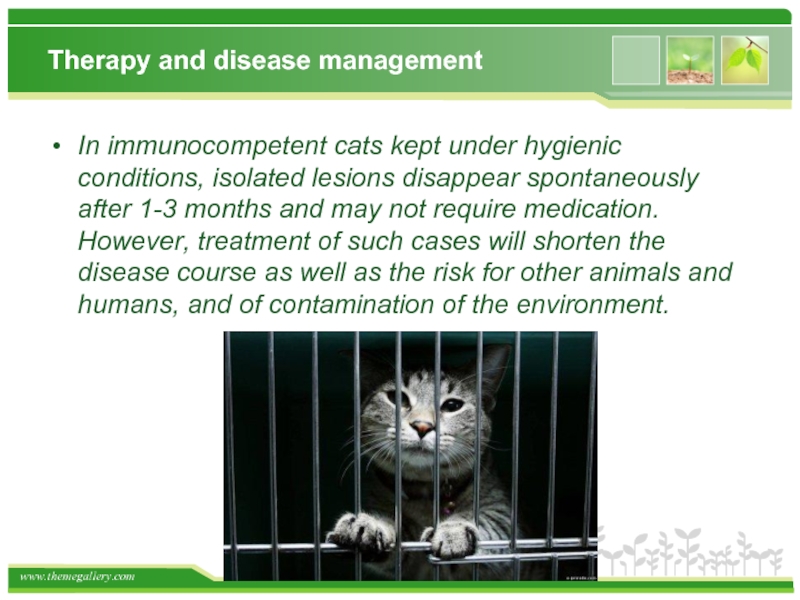
In immunocompetent cats kept under hygienic conditions,
isolated lesions disappear spontaneously after 1-3 months and may not require medication. However, treatment of such cases will shorten the disease course as well as the risk for other animals and humans, and of contamination of the environment.
Слайд 18
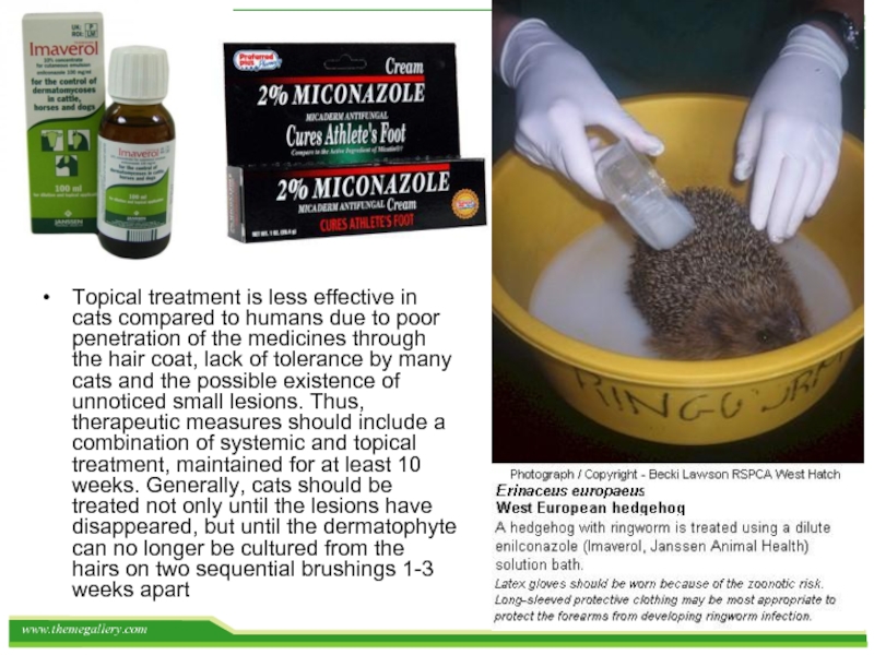
Topical treatment is less effective in cats compared to humans due
to poor penetration of the medicines through the hair coat, lack of tolerance by many cats and the possible existence of unnoticed small lesions. Thus, therapeutic measures should include a combination of systemic and topical treatment, maintained for at least 10 weeks. Generally, cats should be treated not only until the lesions have disappeared, but until the dermatophyte can no longer be cultured from the hairs on two sequential brushings 1-3 weeks apart
Слайд 19Systemic therapy
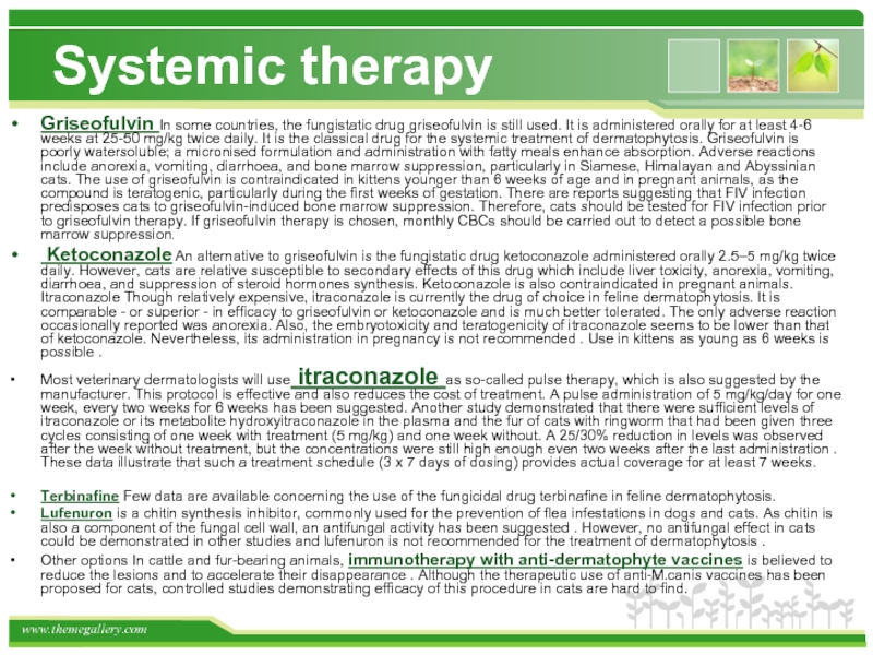
Griseofulvin In some countries, the fungistatic drug griseofulvin is still
used. It is administered orally for at least 4-6 weeks at 25-50 mg/kg twice daily. It is the classical drug for the systemic treatment of dermatophytosis. Griseofulvin is poorly watersoluble; a micronised formulation and administration with fatty meals enhance absorption. Adverse reactions include anorexia, vomiting, diarrhoea, and bone marrow suppression, particularly in Siamese, Himalayan and Abyssinian cats. The use of griseofulvin is contraindicated in kittens younger than 6 weeks of age and in pregnant animals, as the compound is teratogenic, particularly during the first weeks of gestation. There are reports suggesting that FIV infection predisposes cats to griseofulvin-induced bone marrow suppression. Therefore, cats should be tested for FIV infection prior to griseofulvin therapy. If griseofulvin therapy is chosen, monthly CBCs should be carried out to detect a possible bone marrow suppression.
Ketoconazole An alternative to griseofulvin is the fungistatic drug ketoconazole administered orally 2.5–5 mg/kg twice daily. However, cats are relative susceptible to secondary effects of this drug which include liver toxicity, anorexia, vomiting, diarrhoea, and suppression of steroid hormones synthesis. Ketoconazole is also contraindicated in pregnant animals. Itraconazole Though relatively expensive, itraconazole is currently the drug of choice in feline dermatophytosis. It is comparable - or superior - in efficacy to griseofulvin or ketoconazole and is much better tolerated. The only adverse reaction occasionally reported was anorexia. Also, the embryotoxicity and teratogenicity of itraconazole seems to be lower than that of ketoconazole. Nevertheless, its administration in pregnancy is not recommended . Use in kittens as young as 6 weeks is possible .
Most veterinary dermatologists will use itraconazole as so-called pulse therapy, which is also suggested by the manufacturer. This protocol is effective and also reduces the cost of treatment. A pulse administration of 5 mg/kg/day for one week, every two weeks for 6 weeks has been suggested. Another study demonstrated that there were sufficient levels of itraconazole or its metabolite hydroxyitraconazole in the plasma and the fur of cats with ringworm that had been given three cycles consisting of one week with treatment (5 mg/kg) and one week without. A 25/30% reduction in levels was observed after the week without treatment, but the concentrations were still high enough even two weeks after the last administration . These data illustrate that such a treatment schedule (3 x 7 days of dosing) provides actual coverage for at least 7 weeks.
Terbinafine Few data are available concerning the use of the fungicidal drug terbinafine in feline dermatophytosis.
Lufenuron is a chitin synthesis inhibitor, commonly used for the prevention of flea infestations in dogs and cats. As chitin is also a component of the fungal cell wall, an antifungal activity has been suggested . However, no antifungal effect in cats could be demonstrated in other studies and lufenuron is not recommended for the treatment of dermatophytosis .
Other options In cattle and fur-bearing animals, immunotherapy with anti-dermatophyte vaccines is believed to reduce the lesions and to accelerate their disappearance . Although the therapeutic use of anti-M.canis vaccines has been proposed for cats, controlled studies demonstrating efficacy of this procedure in cats are hard to find.
Слайд 20
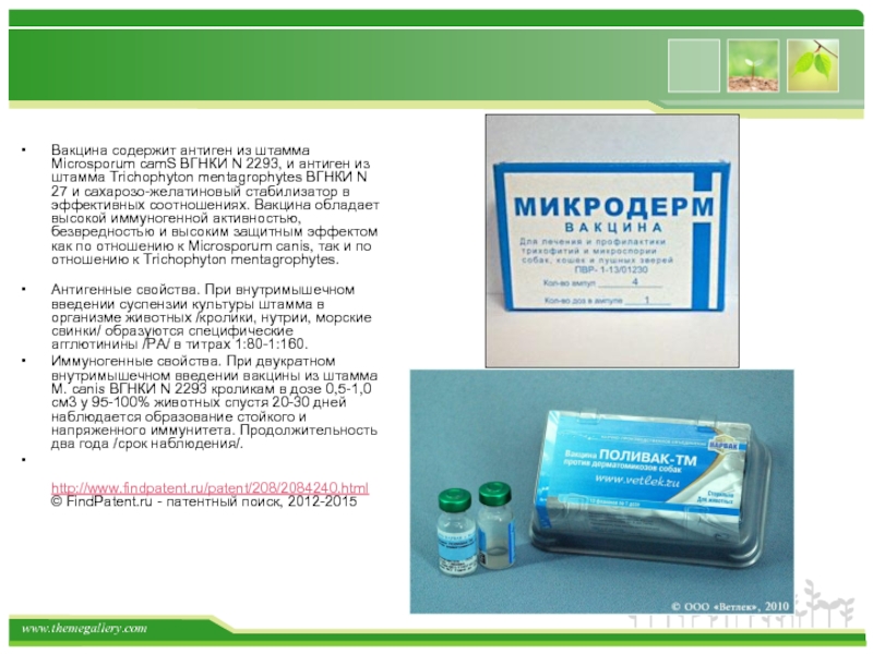
Вакцина содержит антиген из штамма Microsporum camS ВГНКИ N 2293, и
антиген из штамма Trichophyton mentagrophytes ВГНКИ N 27 и сахарозо-желатиновый стабилизатор в эффективных соотношениях. Вакцина обладает высокой иммуногенной активностью, безвредностью и высоким защитным эффектом как по отношению к Microsporum canis, так и по отношению к Trichophyton mentagrophytes.
Антигенные свойства. При внутримышечном введении суспензии культуры штамма в организме животных /кролики, нутрии, морские свинки/ образуются специфические агглютинины /РА/ в титрах 1:80-1:160.
Иммуногенные свойства. При двукратном внутримышечном введении вакцины из штамма M. canis ВГНКИ N 2293 кроликам в дозе 0,5-1,0 см3 у 95-100% животных спустя 20-30 дней наблюдается образование стойкого и напряженного иммунитета. Продолжительность два года /срок наблюдения/.
http://www.findpatent.ru/patent/208/2084240.html © FindPatent.ru - патентный поиск, 2012-2015
