- Главная
- Разное
- Дизайн
- Бизнес и предпринимательство
- Аналитика
- Образование
- Развлечения
- Красота и здоровье
- Финансы
- Государство
- Путешествия
- Спорт
- Недвижимость
- Армия
- Графика
- Культурология
- Еда и кулинария
- Лингвистика
- Английский язык
- Астрономия
- Алгебра
- Биология
- География
- Детские презентации
- Информатика
- История
- Литература
- Маркетинг
- Математика
- Медицина
- Менеджмент
- Музыка
- МХК
- Немецкий язык
- ОБЖ
- Обществознание
- Окружающий мир
- Педагогика
- Русский язык
- Технология
- Физика
- Философия
- Химия
- Шаблоны, картинки для презентаций
- Экология
- Экономика
- Юриспруденция
Hemostasis презентация
Содержание
- 1. Hemostasis
- 2. Hemostasis („hemo”=blood; sta=„remain”) is the stoppage of
- 3. Local vasoconstriction is due to local spasm
- 4. Formation of platelet aggregate Injured blood
- 5. The micrograph shows activated platelets adhering to some damaged cells
- 6. Formation of
- 7. The intrinsic system is more complex
- 8. Extrinsic pathway: 1. When blood comes
- 9. Intrinsic pathway: 2. Exposed collagen activates Hageman
- 10. Stage I: Formation of prothrombin activator 3.
- 12. Stage II: conversion of prothrombin to thrombin
- 13. Stage III: conversion of fibrinogen to fibrin
- 14. Calcium ions Are required for promotion and
- 15. Ca2+ Ca2+ Christmas
- 18. Fibrinolysis
- 19. Clot Dissolution Plasmin is formed from plasminogen
- 21. Anticoagulants Hirudo medicinalis produce Hirudin that inhibits Thrombin
- 22. Anticoagulants Although tissue breakdown and platelets destruction
- 23. Natural anticoagulants Antithrombin III – inhibits factor
- 24. Abnormalities of hemostasis
- 25. Thrombocytopenia Severe reduction in the number of
- 26. Thrombocytopenia Lethal when PLTs
- 27. Hepatic failure Most of the clotting factors are formed in the liver Subconjunctival hemorrhage
- 28. Disseminated intravascular coagulation (DIC) Widespread coagulation →
- 29. Hemophilia A (lack of F VIII) and
- 30. Hemophilia A (lack of F VIII;
- 31. Son of the last Tsar of Russia – Aleksy Romanow suffered from Hemophilia A
- 32. Tests of coagulation
- 33. "Intrinsic" and "extrinsic" coagulation pathways N: 9.9
- 34. Prothrombin time (PT) test – norm 11
- 35. International Normalised Ratio (INR) The result for
- 36. Activated Partial Thromboplastin Time test (aPTT) –
- 37. Thrombin time (TT) – norm: 14-15 sec
- 38. Selected causes of abnormal coagulation tests
- 39. "Intrinsic" and "extrinsic" coagulation pathways N: 9.9
- 40. Whole blood clotting time The time
- 41. Whole blood clotting time
- 42. Bleeding time This is a test
- 43. Bleeding time
- 44. Abnormal Bleeding Time Prolonged bleeding time may
- 45. http://www.medicine.mcgill.ca/physio/vlab212D/bloodlab/images/clottime5.mpg
- 47. The new model of haemostasis
- 48. Injury of vessels wall leads to contact
- 49. 2. Amplification phase Activated platelets bind FVa, FVIIIa and FIXa
- 50. 3. Propagation phase The “thrombin
- 51. Summary: Haemostasis starts with the interaction
- 52. NovoSeven® Mode of Action Eptacog alfa (activated)
- 53. Conclusion: • In high doses rFVIIa binds
- 54. Prescribing Information NovoSeven® Eptacog alfa (activated)
- 55. A 35-year-old man complains of chronic physical
- 56. Laboratory findings revealed the following:
- 57. Case history questions: What general medical condition
- 58. Answers: Anemia A reduction in oxygen-carrying capacity

Слайд 2Hemostasis („hemo”=blood; sta=„remain”) is the stoppage of bleeding, which is vitally
Following an injury to blood vessels several actions may help prevent blood loss, including:
Formation of a clot
 is the stoppage of bleeding, which is vitally important when blood vessels")
Слайд 3Local vasoconstriction
is due to local spasm of the smooth muscle (symp.
can be maintained by platelet vasoconstrictors
can be maintained by")
Слайд 4Formation of
platelet aggregate
Injured blood vessel releases ADP, which attracts platelets
PLT comming in contact with exposed collagen release: serotonin, ADP, TXA2, which accelerate vasoconstriction and causes PLT to swell and become more sticky
PLT comming in contact")

Слайд 6Formation of
In the formation of the clot, an enzyme called thrombin converts fibrinogen into insoluble protein, fibrin
Fibrin aggregates to form a meshlike network at the site of vascular damage

Слайд 7
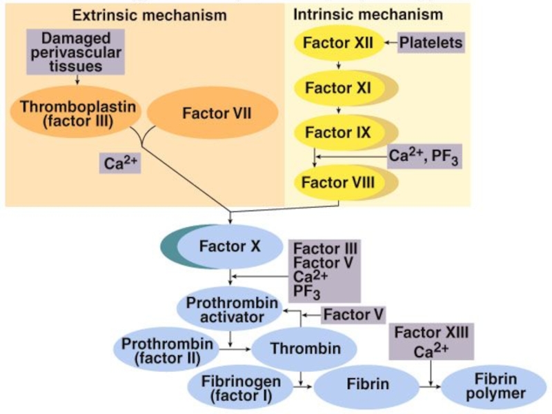
The intrinsic system is more complex and present only in „higher”
The complex sequence of events that produce fibrin are divided into three stages
Coagulation mechanism is composed of an extrinsic and intrinsic pathway, which eventually merge into one

Слайд 8Extrinsic pathway:
1. When blood comes in contact with injured tissue –
Stage I: Formation of prothrombin activator
Ca2+
Stuart factor
Anti- hemophilic factor
Christmas factor
")
Слайд 9Intrinsic pathway:
2. Exposed collagen activates Hageman factor (F XII). Activated F
Stage I: Formation of prothrombin activator
Ca2+
Christmas factor
Anti- hemophilic factor
Stuart factor
. Activated F XII activates plasma enzyme")
Слайд 10Stage I: Formation of prothrombin activator
3. Common pathway:
Activated F X in
Ca2+
Christmas factor
Anti- hemophilic factor
Stuart factor

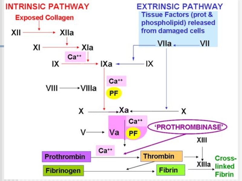
Слайд 12Stage II: conversion of prothrombin to thrombin
Prothrombin – inactive precursor of
In the presence of prothrombin activator and Ca2+ prothrombin is converted to thrombin
Thrombin itself increases its own rate of formation (positive feedback mechanism)
Ca2+

Слайд 13Stage III: conversion of fibrinogen to fibrin
Fibrinogen – plasma protein produced
Thrombin converts fibrinogen to fibrin
Thrombin also activates fibrin-stabilizing factor (F XIII), which in the presence of Ca2+, stabilizes the fibrin polymer through covalent bonding of fibrin monomers
fibrin-stabilizing factor

Слайд 14Calcium ions
Are required for promotion and acceleration of almost all blood
Except: activation of XII and XI (intrinsic mechanism)
Ca2+
http://www.mhhe.com/biosci/esp/2002_general/Esp/folder_structure/tr/m1/s7/trm1s7_3.htm


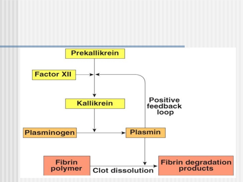
Слайд 19Clot Dissolution
Plasmin is formed from plasminogen - enzyme called activator (e.g.
Plasmin as an enzyme is involved in breaking down fibrin into soluble fragments (fibrinolysis)
Plasminogen Plasmin
Activator (e.g. t-PA)
Fibrin soluble fragments
Plasminogen may be produced by eosinophils


Слайд 22Anticoagulants
Although tissue breakdown and platelets destruction are normal events in the
the amounts of procoagulants released are very small
natural anticoagulants are present (Antithrombin III, Heparin, Antithromboplastin, Protein C and S, fibrin fibers)

Слайд 23Natural anticoagulants
Antithrombin III – inhibits factor X and thrombin
Heparin from
Antithromboplastin (inhibits „tissue factors” – tissue thromboplastins)
Protein C and S – activated by thrombin; degrade factor Va and VIIIa


Слайд 25Thrombocytopenia
Severe reduction in the number of PLTs - thrombocytopenia
this causes spontaneous
in the skin - reddish-purple blotchy rash
it may result from:
decreased production (toxins, radiation, infection, leukemias)
increased destruction (autoimmune processes)
increased PLTs consumption (DIC)
Hemorrhagic spots (petechiae)


Слайд 27Hepatic failure
Most of the clotting factors are formed in the liver
Subconjunctival

Слайд 28Disseminated intravascular coagulation (DIC)
Widespread coagulation →
It may result from:
bacterial infections (endothelial damage)
disseminated cancers (release of procoagulants)
complications of pregnancy
severe catabolic states
Disseminated cervical cancer metastases (PET imaging)
Widespread coagulation →")
Слайд 29Hemophilia A (lack of F VIII) and B (lack of F
Von Willebrand’s disease – loss of large component of fVIII
 and B (lack of F IX) are transmitted genetically")
Слайд 30Hemophilia A
(lack of F VIII; 85%)
Spontaneous or traumatic subcutaneous bleeding
Blood
Bleeding in the mouth, lips, tongue
Bleeding to the joints, CNS, gastrointestinal tract
Mild hemophilia after injection in buttock
Spontaneous or traumatic subcutaneous bleedingBlood in the urineBleeding in")


Слайд 33"Intrinsic" and "extrinsic" coagulation pathways
N: 9.9 – 13 sec
Activated Partial Thromboplastin
N: 25-35 sec
Prothrombin Time
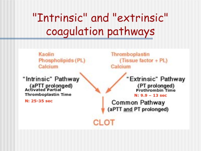
Слайд 34Prothrombin time (PT) test – norm 11 -15 sec evaluates extrinsic system
prolonged PT indicates a deficiency in any of factors VII, X, V, prothrombin (factor II), or fibrinogen (factor I).
Prolonged PT:
- a vitamin K deficiency (vitamin K is a co-factor in the synthesis of functional factors II (prothrombin), VII, IX and X)
- liver disease
Warfarin therapy
DIC
excesive heparin
 test – norm 11 -15 sec evaluates extrinsic system (VII, X, V,")
Слайд 35International Normalised Ratio (INR)
The result for the PT is expressed as
Correction factor (International Sensitivity Index) is applied to the prothrombin ratio and the result issued as INR.
Therapeutic interval: Therapeutic interval for oral anticoagulant therapy: 2.0-4.5.
Application: Monitoring oral anticoagulant therapy (eg. Warfarin);
note that heparin will not prolong INR (heparinase is included within the INR reagent)!!!!!!!!!!!!! For heparin therapy we monitor aPTT and/or aPTT ratio
The result for the PT is expressed as a ratio (prothrombin clotting")
Слайд 36Activated Partial Thromboplastin Time test (aPTT) – norm: 25-35 s; evaluates
an isolated prolongation of the aPTT (PT normal) suggests deficiency of factor VIII, IX, XI or XII
prolongation of both the APTT and PT suggests factor X, V, II or I (fibrinogen) deficiency, all of which are rare
aPTT is normal in factor VII deficiency (PT prolonged) and factor XIII deficiency
Most common case of prolonged aPTT – heparin!!!
 – norm: 25-35 s; evaluates intrinsic system (VIII, IX,")
Слайд 37Thrombin time (TT) – norm: 14-15 sec
Prolonged TT:
Heparin (much more sensitive
Hypofibrinogenemia
 – norm: 14-15 secProlonged TT:Heparin (much more sensitive to heparin than aPTT)Hypofibrinogenemia")

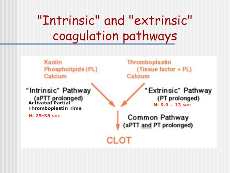
Слайд 39"Intrinsic" and "extrinsic" coagulation pathways
N: 9.9 – 13 sec
Activated Partial Thromboplastin
N: 25-35 sec
Слайд 40Whole blood
clotting time
The time taken for blood to clot mainly
The surface of the glass tube initiates the clotting process. This test is sensitive to the factors involved in the intrinsic pathway
The expected range for clotting time is 4-10 mins.

Слайд 41Whole blood clotting time
Clean the tip of the finger with an alcohol
Prick the finger tip with an automatic lancet
Note the time when blood first appears on the skin
Touch the tube to the drop of blood
Break gently 1cm of the tube at the end of 2 min, and every 30 sec these after
When fibrin is formed between the two broken pieces of tube the coagulation or clotting time is noted

Слайд 42Bleeding time
This is a test that measures
This test is useful for detecting bleeding tendencies
The bleeding stops within 1 to 9 minutes. This may vary from lab to lab, depending on how the test is measured
Using the ear lobe method, a normal bleeding time is between 1 and 4 minutes.

Слайд 43Bleeding time
Clean the earlobe with an alcohol
Prick the earlobe with an automatic lancet
Note the time when blood first appears on the skin
After half a minute (30sec) place the edge of the filter paper on the top of the drop of blood.
Perform the operation at half minute (30 sec) interval
The end point or bleeding time is the first half minute when no blood is seen on the filter paper.

Слайд 44Abnormal Bleeding Time
Prolonged bleeding time may indicate:
A vascular (blood vessel)
A platelet function defect (see platelet aggregation)
platelets count defect (low platelets)
Drugs that may increase times include dextran, indomethacin, and salicylates (including aspirin).
 defect A platelet function")


Слайд 48Injury of vessels wall
leads to contact
between blood and
subendothelial cells
FXa
The complex between
TF and FVIIa activates
FIX and FX
Tissue factor (TF) is
exposed and binds to
FVIIa or FVII which
is subsequently
converted to FVIIa
1. Initiation phase


Слайд 51Summary:
Haemostasis starts with the interaction between TF and FVIIa on
The small amount of thrombin generated during the amplification phase activates platelets locally on whose surface the subsequent reactions take place.
The resulting thrombin burst results in the formation of a stable clot.

Слайд 52NovoSeven® Mode of Action
Eptacog alfa (activated)
The thrombin burst leads
to the
The thrombin burst leads to the formation of a stable clot")
Слайд 53Conclusion:
• In high doses rFVIIa binds to the surface of the

Слайд 54Prescribing Information
NovoSeven® Eptacog alfa (activated) Abbreviated Prescribing Information: NovoSeven [Recombinant Coagulation
Serious adverse reactions include: Arterial thrombotic events (such as myocardial infarction or ischaemia, cerebrovascular disorders and bowel infarction); venous thrombotic events (such as thrombophlebitis, deep vein thrombosis and pulmonary embolism). In the vast majority of cases patients were predisposed to such events. No spontaneous reports of anaphylactic reactions, but patients with a history of allergic reaction should be carefully monitored. No reports of antibodies against FVII in haemophilia A or B patients. Isolated cases of FVII-deficient patients developing antibodies against FVII reported after treatment with NovoSeven. These patients previously treated with human plasma and/or plasma derived FVII. Monitor FVII deficient patients for FVII antibodies. One case angioneurotic oedema reported in patient with Glanzmann’s thrombasthenia after administration of NovoSeven. Marketing Authorisation numbers: NovoSeven 60 KIU EU/1/96/006/001 NovoSeven 120 KIU EU/1/96/006/002 NovoSeven 240 KIU EU/1/96/006/003 Legal Category: POM Basic NHS Price: NovoSeven 1.2 mg £664.72 NovoSeven 2.4 mg £1329.44 NovoSeven 4.8 mg £2658.88 Further information: Full prescribing information can be obtained from: Novo Nordisk Limited Broadfield Park Brighton Road Crawley West Sussex RH11 9RT Tel: 01293 613555 Fax: 01293 613535 Date of preparation: May 2004 Ref N7/03/039a
 Abbreviated Prescribing Information: NovoSeven [Recombinant Coagulation Factor VIIa (rFVIIa)] Presentation:")
Слайд 55A 35-year-old man complains of chronic physical fatigue, which began 3-4


Слайд 57Case history questions:
What general medical condition is suggested by the person’s
What fundamental change in function of blood related to the red blood cells could simultaneously affect the function of several systems (cardiovascular, respiratory, gastrointestinal, and others)?
What specific diagnosis is supported by the laboratory findings?
How could the stool be related to the laboratory findings?

Слайд 58Answers:
Anemia
A reduction in oxygen-carrying capacity of the blood and thus a
An iron defficiency anemia
Most cases of iron-defficiency anemia result from internal blood loss. Dark, tarry loose stools suggest bleeding from the gastrointestinal tract and warrant further tests to determine the exact cause