- Главная
- Разное
- Дизайн
- Бизнес и предпринимательство
- Аналитика
- Образование
- Развлечения
- Красота и здоровье
- Финансы
- Государство
- Путешествия
- Спорт
- Недвижимость
- Армия
- Графика
- Культурология
- Еда и кулинария
- Лингвистика
- Английский язык
- Астрономия
- Алгебра
- Биология
- География
- Детские презентации
- Информатика
- История
- Литература
- Маркетинг
- Математика
- Медицина
- Менеджмент
- Музыка
- МХК
- Немецкий язык
- ОБЖ
- Обществознание
- Окружающий мир
- Педагогика
- Русский язык
- Технология
- Физика
- Философия
- Химия
- Шаблоны, картинки для презентаций
- Экология
- Экономика
- Юриспруденция
Heart pathology. (Subject 13) презентация
Содержание
- 1. Heart pathology. (Subject 13)
- 2. Lecture Plan Signs and symptoms of MI Cardiogenic shock Arrhythmia classification Characteristic of arrhythmia’s types
- 3. Signs and symptoms of MI Chest pain
- 4. Signs and symptoms of MI A wide
- 5. Signs and symptoms of MI Enzymes and
- 6. Reperfusion of MI circulation brings neutrophils
- 7. Cardiogenic shock Cardiogenic shock is a
- 8. Cardiogenic shock symptoms Anxiety, restlessness, altered mental
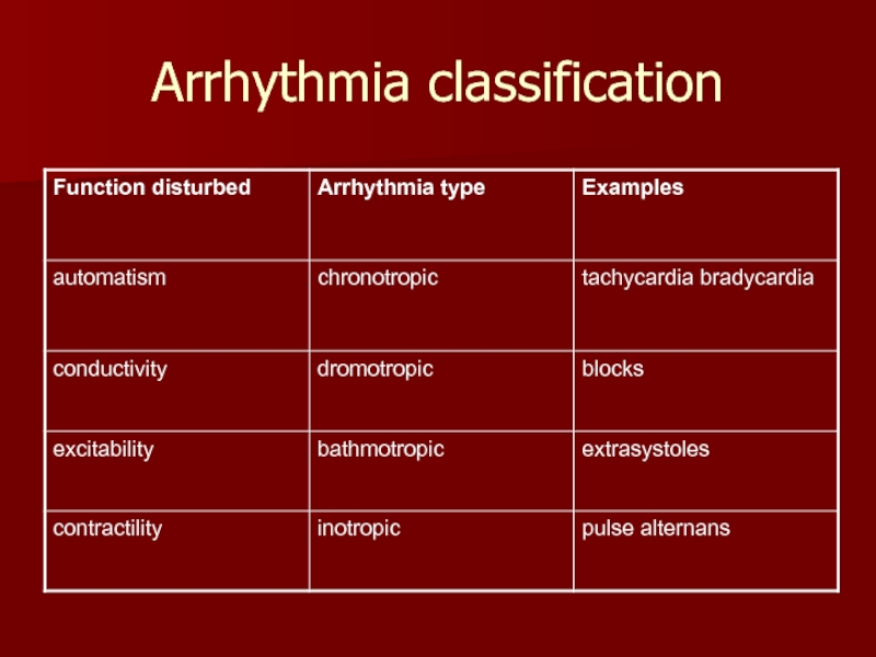
- 9. Arrhythmia classification
- 10. Pathology of automatism Sinus tachycardia –
- 11. Pathology of automatism Sinus bradycardia –
- 12. Pathology of automatism Sinus arrhythmia fluctuation of
- 13. Conduction abnormalities Sino-atrial block is characterized
- 14. Atrioventricular block Atrioventricular block is the blockage
- 15. Atrioventricular block 2nd degree AV block- some
- 16. Atrioventricular block 3rd degree AV block (complete
- 17. Bundle branch block Bundle branch block
- 18. Pathology of excitability Pathology of excitability
- 19. Sinus extrasystole Sinus extrasystole originates in
- 20. Atrial ectopic beat Atrial ectopic beats
- 21. Premature junctional contractions Ectopic beat originate in
- 22. Ventricular ectopic beat wide QRS-complex (above 0.12 s), long compensatory interval (2RR)
- 23. Paroxysmal ectopic tachycardia Paroxysmal atrial tachycardia
- 24. Paroxysmal ectopic tachycardia Paroxysmal ventricular tachycardia ≤
- 25. Disorders of hemodynamic in the pathology of
- 26. Atrial fibrillation and flutter Atrial fibrillation -
- 27. Reasons of atrial fibrillation Re-entry phenomenon -
- 28. Ventricular fibrillation Ventricular fibrillation irregular ventricular rate
- 29. Defibrillation of the heart Defibrillation – brings
- 30. Pathology of contractility Pulsus alternans – alternation
Слайд 2Lecture Plan
Signs and symptoms of MI
Cardiogenic shock
Arrhythmia classification
Characteristic of arrhythmia’s types
Слайд 3Signs and symptoms of MI
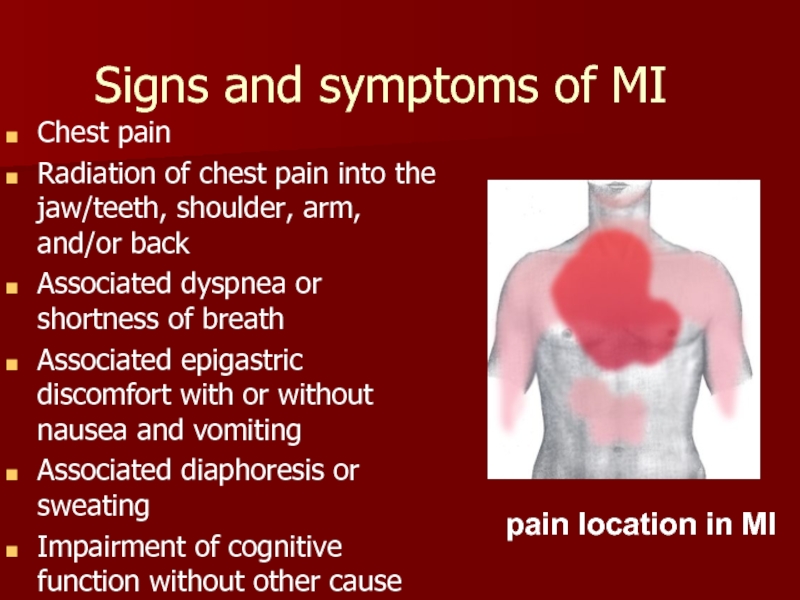
Chest pain
Radiation of chest pain into the
Associated dyspnea or shortness of breath
Associated epigastric discomfort with or without nausea and vomiting
Associated diaphoresis or sweating
Impairment of cognitive function without other cause
pain location in MI
Слайд 4Signs and symptoms of MI
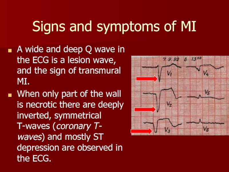
A wide and deep Q wave in
When only part of the wall is necrotic there are deeply inverted, symmetrical T-waves (coronary T- waves) and mostly ST depression are observed in the ECG.
Слайд 5Signs and symptoms of MI
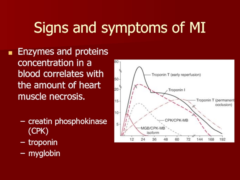
Enzymes and proteins concentration in a blood
creatin phosphokinase (CPK)
troponin
myglobin
Слайд 6Reperfusion of MI
circulation brings neutrophils to re-perfused tissues that release
reperfusion brings a massive influx of Ca++ which leads to activation of enzymes progressive destruction of all cell structures.
Слайд 7Cardiogenic shock
Cardiogenic shock is a severe reduction of cardiac output
The pulmonary capillary wedge pressure is normal or elevated in contrast to other types of shock (blood loss or vasodilatation).
The cardiac pump do not get rid of the blood volume received and it is therefore accumulated in venous system
The lower part of a body is filled with blood in distensible vessels, and the upper part of the body is pale.
Слайд 8Cardiogenic shock symptoms
Anxiety, restlessness, altered mental state
Hypotension
A rapid, weak, thready pulse
Cool, clammy, and mottled skin (cutis marmorata)
Distended jugular veins
Oliguria (low urine output)
Rapid and deep respirations (hyperventilation)
Fatigue
Слайд 10Pathology of automatism
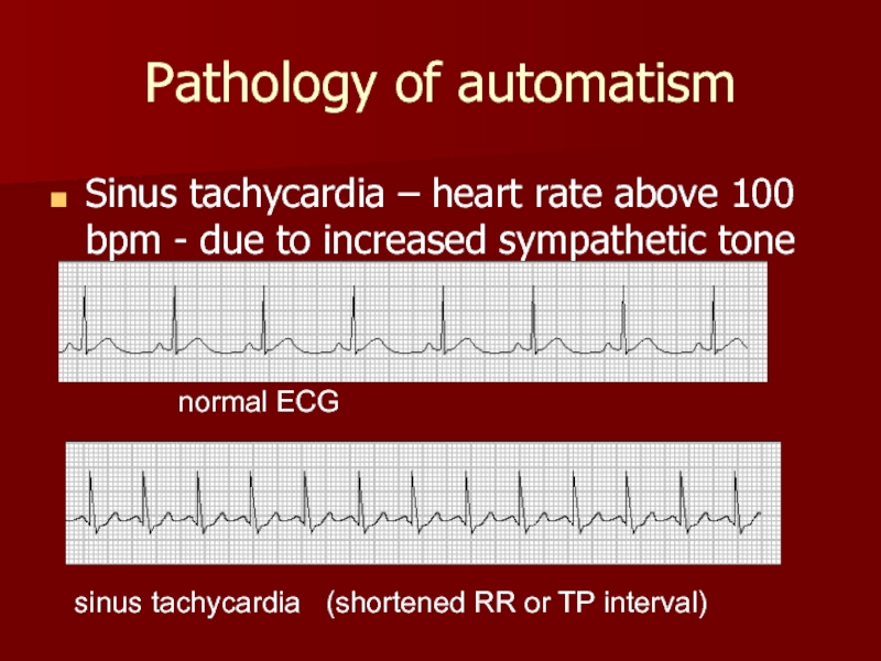
Sinus tachycardia – heart rate above 100 bpm
normal ECG
sinus tachycardia (shortened RR or TP interval)
Слайд 11Pathology of automatism
Sinus bradycardia – less than 60 bpm due
normal ECG
sinus bradycardia (increased RR or TP interval)
Слайд 12Pathology of automatism
Sinus arrhythmia fluctuation of the vagal tone due to
normal ECG
Expiration
Inspiration
Слайд 13Conduction abnormalities
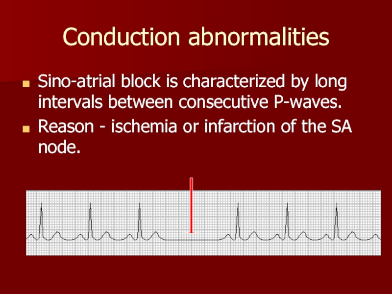
Sino-atrial block is characterized by long intervals between consecutive
Reason - ischemia or infarction of the SA node.
Слайд 14Atrioventricular block
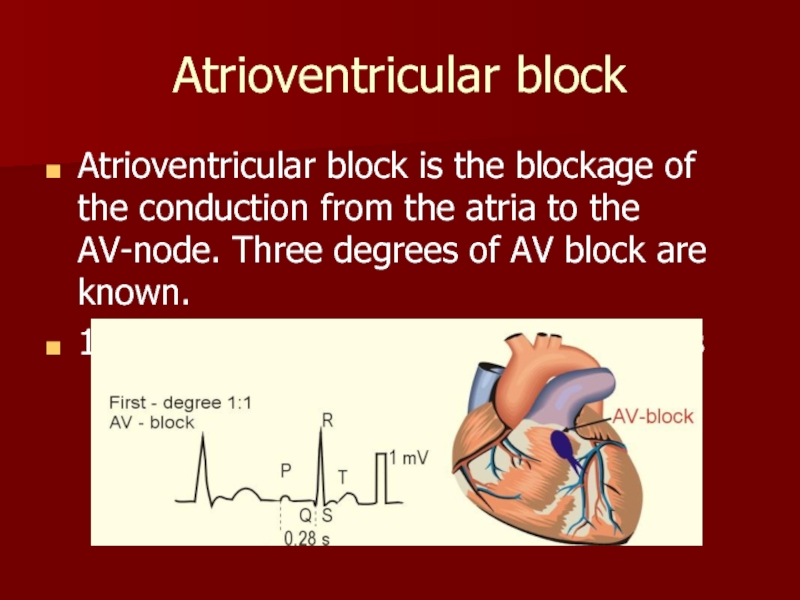
Atrioventricular block is the blockage of the conduction from the
1st degree AV block: PQ - above 0.2 s
Слайд 15Atrioventricular block
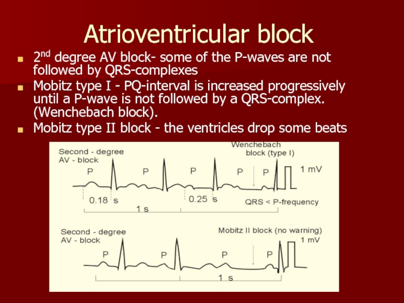
2nd degree AV block- some of the P-waves are not
Mobitz type I - PQ-interval is increased progressively until a P-wave is not followed by a QRS-complex. (Wenchebach block).
Mobitz type II block - the ventricles drop some beats
Слайд 16Atrioventricular block
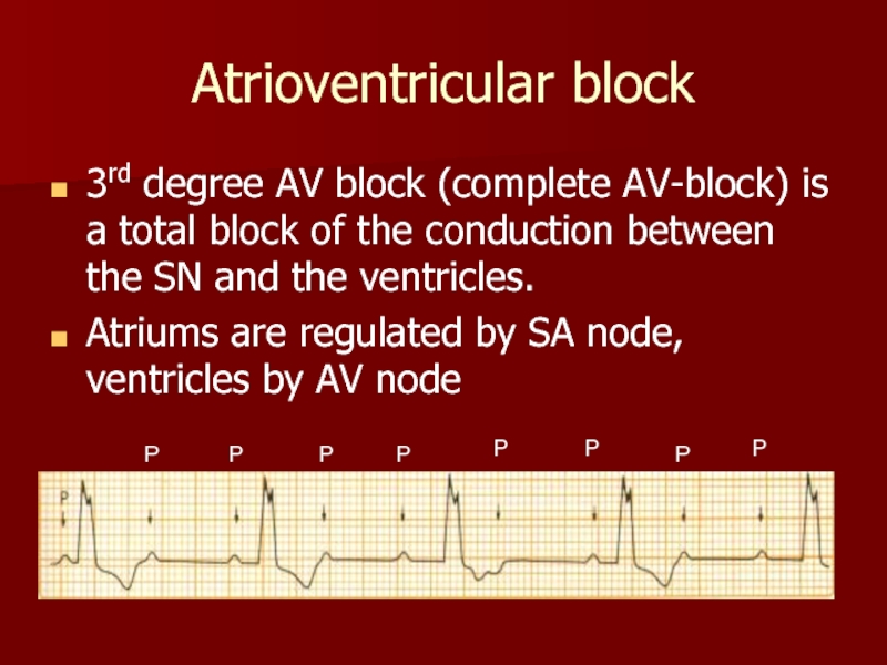
3rd degree AV block (complete AV-block) is a total block
Atriums are regulated by SA node, ventricles by AV node
P
P
P
P
P
P
P
P
Слайд 17Bundle branch block
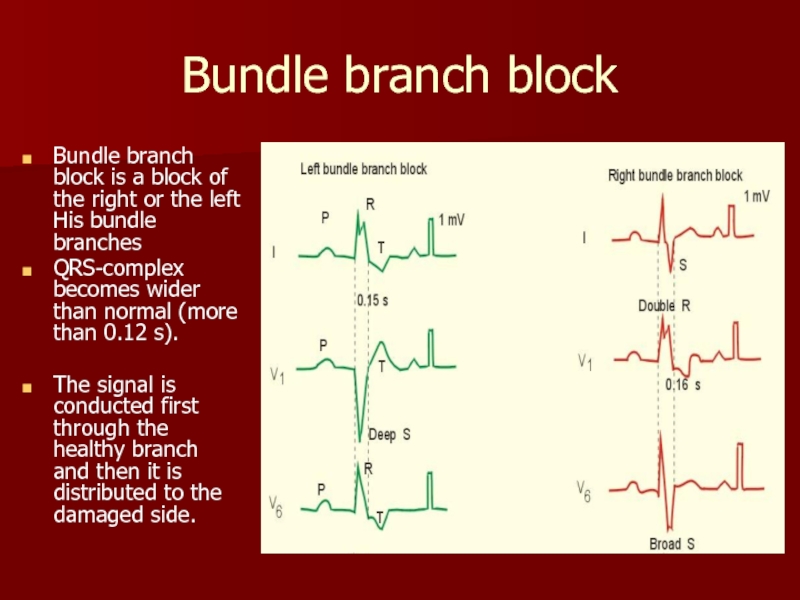
Bundle branch block is a block of the
QRS-complex becomes wider than normal (more than 0.12 s).
The signal is conducted first through the healthy branch and then it is distributed to the damaged side.
Слайд 18Pathology of excitability
Pathology of excitability is usually manifested with ectopic
extrasystole (premature contraction, ectopic beat)
paroxysmal tachycardia
fibrillation.
Reasons: ischaemia, mechanical or chemical stimuli, metabolic disturbances..
Слайд 19Sinus extrasystole
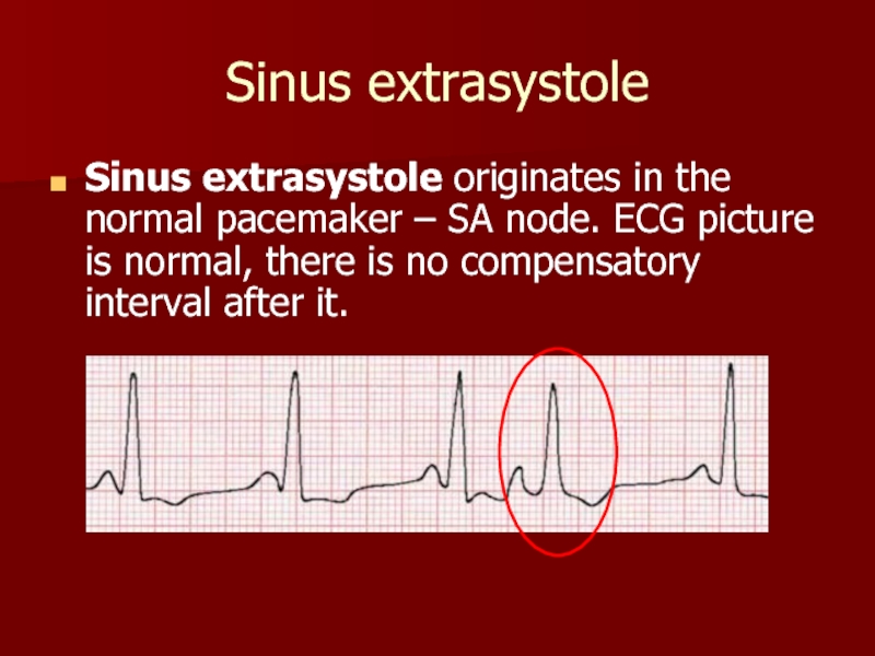
Sinus extrasystole originates in the normal pacemaker – SA
Слайд 20Atrial ectopic beat
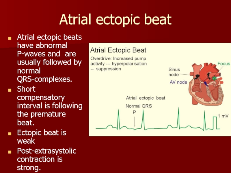
Atrial ectopic beats have abnormal P-waves and are
Short compensatory interval is following the premature beat.
Ectopic beat is weak
Post-extrasystolic contraction is strong.
Слайд 21Premature junctional contractions
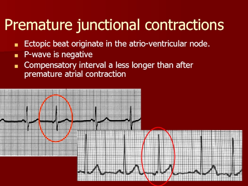
Ectopic beat originate in the atrio-ventricular node.
P-wave is
Compensatory interval a less longer than after premature atrial contraction
Слайд 23Paroxysmal ectopic tachycardia
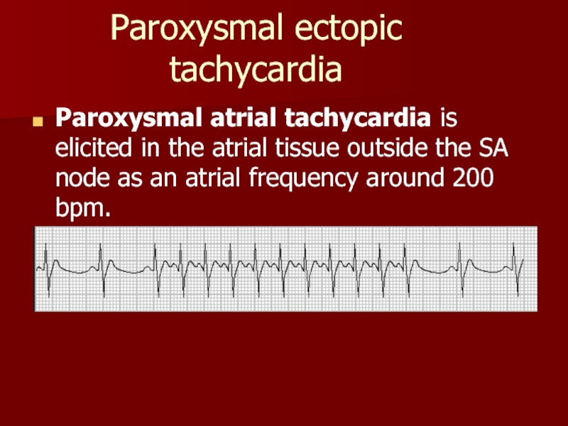
Paroxysmal atrial tachycardia is elicited in the atrial
Слайд 24Paroxysmal ectopic tachycardia
Paroxysmal ventricular tachycardia ≤ 120 bpm
P-waves are absent
QRS-complexes are
Слайд 25Disorders of hemodynamic in the pathology of excitability
Single extrasystole clinically manifests
Plural extrasystoles can seriously violate the hemodynamic:
extrasystoles appear in different phases of cardiac cycle - so they are ineffective in hemodynamic
Myocardium can’t react to the normal impulse during compensatory pause following extrasystole
Слайд 26Atrial fibrillation and flutter
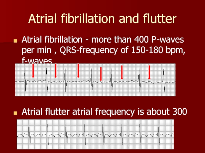
Atrial fibrillation - more than 400 P-waves per
Atrial flutter atrial frequency is about 300 bpm, sawtooth-like P-waves
Слайд 27Reasons of atrial fibrillation
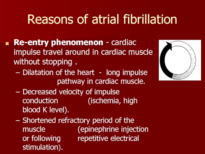
Re-entry phenomenon - cardiac impulse travel around in
Dilatation of the heart - long impulse pathway in cardiac muscle.
Decreased velocity of impulse conduction (ischemia, high blood K level).
Shortened refractory period of the muscle (epinephrine injection or following repetitive electrical stimulation).
Слайд 28Ventricular fibrillation
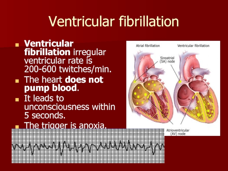
Ventricular fibrillation irregular ventricular rate is 200-600 twitches/min.
The heart
It leads to unconsciousness within 5 seconds.
The trigger is anoxia.
Слайд 29Defibrillation of the heart
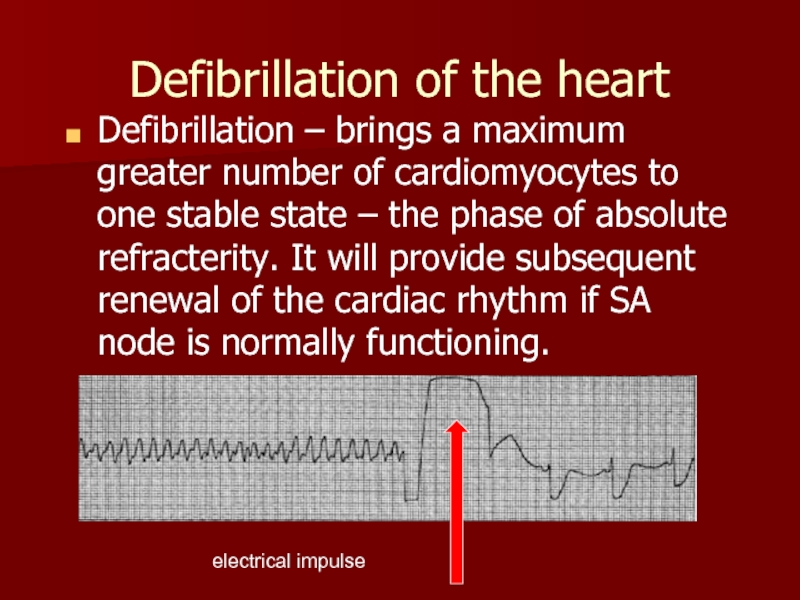
Defibrillation – brings a maximum greater number of
electrical impulse
Слайд 30Pathology of contractility
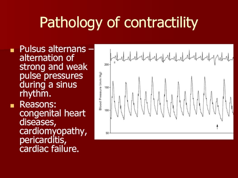
Pulsus alternans – alternation of strong and weak pulse
Reasons: congenital heart diseases, cardiomyopathy, pericarditis, cardiac failure.
