- Главная
- Разное
- Дизайн
- Бизнес и предпринимательство
- Аналитика
- Образование
- Развлечения
- Красота и здоровье
- Финансы
- Государство
- Путешествия
- Спорт
- Недвижимость
- Армия
- Графика
- Культурология
- Еда и кулинария
- Лингвистика
- Английский язык
- Астрономия
- Алгебра
- Биология
- География
- Детские презентации
- Информатика
- История
- Литература
- Маркетинг
- Математика
- Медицина
- Менеджмент
- Музыка
- МХК
- Немецкий язык
- ОБЖ
- Обществознание
- Окружающий мир
- Педагогика
- Русский язык
- Технология
- Физика
- Философия
- Химия
- Шаблоны, картинки для презентаций
- Экология
- Экономика
- Юриспруденция
General course of syphilis. Primary syphilis secondary syphslis презентация
Содержание
- 1. General course of syphilis. Primary syphilis secondary syphslis
- 2. Theoretical part Etiology of syphilis. Diagnostic laboratory
- 3. Classification of syphilis Primary, seronegative syphilis –
- 4. Classification of syphilis Latent syphilis – syphilis
- 5. Clinical features of primary syphilis. After
- 6. Clinical features of primary syphilis. The extragenital
- 7. Clinical features of primary syphilis. Regional scleradenitis
- 8. Atypical Chancres Chancre-amygdalitis is characterized by
- 9. Atypical Chancres Indurative swelling as a manifestation
- 10. Complications of hard chancre Balanitis is the
- 11. Complications of hard chancre Balanoposthitis may lead
- 12. Complications of hard chancre As a result,
- 13. Complications of hard chancre An attempt to
- 14. Complications of hard chancre The development of
- 15. Complications of hard chancre The scab covers
- 16. Secondary syphilis Secondary syphilis develops 2.5-3, rarely
- 17. Secondary Siphilis Characteristic signs of secondary syphilis
- 18. Secondary syphilis Macular syphilis, or syphilitic roseola.
- 19. Diagnosis of macular syphilid (roseola) is
- 20. Papular syphilid is often a sign
- 21. Secondary syphilis Papular syphilid
- 22. The following clinical varieties of papular
- 23. Secondary syphilis - Papular syphilid
- 24. Pustular syphilid. Pustular syphilid occurs rarely
- 25. Syphilitic alopecia or calvities Alopecia areata:
- 26. Syphilitic leukoderma, or pigmentary syphilid (leukoderma syphiliticum)
Слайд 2Theoretical part
Etiology of syphilis. Diagnostic laboratory tests for Treponema pallidum
The
basic method in the study of T. pallidum is the dark-field examination, which gives information about the morphology and the movement of the living pathogen.
The materiel for the examination is the tissue fluid, which is taken carefully with the help of bacterial-loop from the surface of the ulcer or erosion cleaned with sterilized isotonic solution. In case of epithelization or cicatrization, a tissue from the local lymphatic node is taken for the examination.
T. pallidum should be differentiated from T. vulgaris, which lives in the mouth cavity and found on genitals.
Conditions and ways of transmission of syphilis. Infection of syphilis takes place through direct and indirect contacts.
Direct contact:
sexual intercourse: 99%;
b) kiss, bite;
c) contact with the uncovered skin of the medical staff , close domestic contacts.
Indirect contact: Transmission of pathogen through the objects of domestic use. Such way of transmission is very rare, as the T. pallidum is a parasite of the tissue and cannot live in the environment for a long time.
Слайд 3Classification of syphilis
Primary, seronegative syphilis – syphilis I seronegativa.
Primary, seropositive syphilis
– syphilis I seropositiva.
Primary latent syphilis – syphilis I latens. This diagnosis is made when the treatment is begun in the primary period of the disease in the absence of subsequent clinical manifestations of syphilis.
Secondary fresh syphilis – syphilis II recens.
Secondary recurrent syphilis – syphilis II recidiva.
Secondary latent syphilis – syphilis II latens. It is diagnosed in patients whose treatment was begun in the secondary fresh or recurrent period in the absence of clinical manifestations of syphilis at the given time.
Tertiary active syphilis – syphilis III activa.
Tertiary latent syphilis – syphilis III latens. This diagnosis is made in patients who have no clinical manifestations of the disease but revealed active manifestations of the tertiary period in the past.
Слайд 4Classification of syphilis
Latent syphilis – syphilis latens:
Early latent syphilis – syphilis
latens praecox;
Late latent syphilis – syphilis latens tarda. This diagnosis is made in cases with no clinical manifestations of the disease, but with positive serological tests.
Early congenital syphilis – syphilis congenita praecox: congenital syphilis of infants (under 1 year of age) and in very young children (from 1 to 4 years old).
Late congenital syphilis – syphilis congenita tarda.
Late congenital syphilis – syphilis congenita latens.
Visceral syphilis (indicating the involved organ).
Syphilis of the nervous system.
Tabes dorsalis.
General paresis – paralysis progressiva.
Слайд 5Clinical features of primary syphilis.
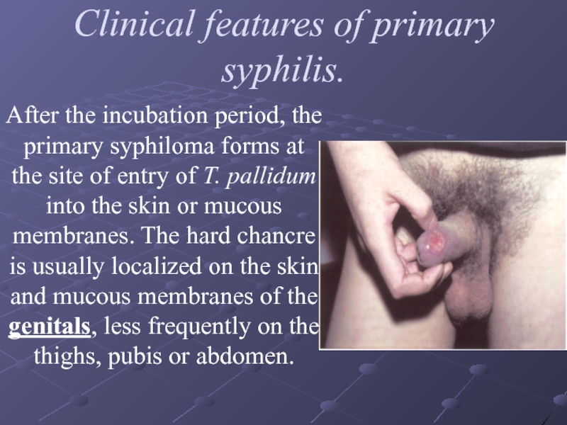
After the incubation period, the primary
syphiloma forms at the site of entry of T. pallidum into the skin or mucous membranes. The hard chancre is usually localized on the skin and mucous membranes of the genitals, less frequently on the thighs, pubis or abdomen.
Слайд 6Clinical features of primary syphilis.
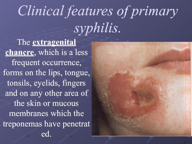
The extragenital chancre, which is a less
frequent occurrence, forms on the lips, tongue, tonsils, eyelids, fingers and on any other area of the skin or mucous membranes which the treponemas have penetrated.
Слайд 7Clinical features of primary syphilis.
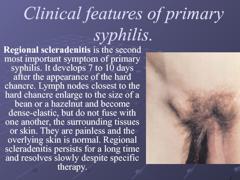
Regional scleradenitis is the second most important
symptom of primary syphilis. It develops 7 to 10 days after the appearance of the hard chancre. Lymph nodes closest to the hard chancre enlarge to the size of a bean or a hazelnut and become dense-elastic, but do not fuse with one another, the surrounding tissues or skin. They are painless and the overlying skin is normal. Regional scleradenitis persists for a long time and resolves slowly despite specific therapy.
Слайд 8Atypical Chancres
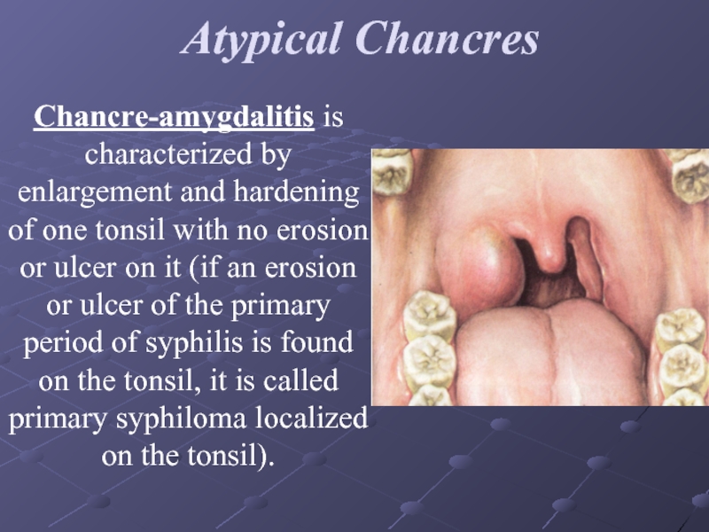
Chancre-amygdalitis is characterized by enlargement and hardening of one
tonsil with no erosion or ulcer on it (if an erosion or ulcer of the primary period of syphilis is found on the tonsil, it is called primary syphiloma localized on the tonsil).
Слайд 9Atypical Chancres
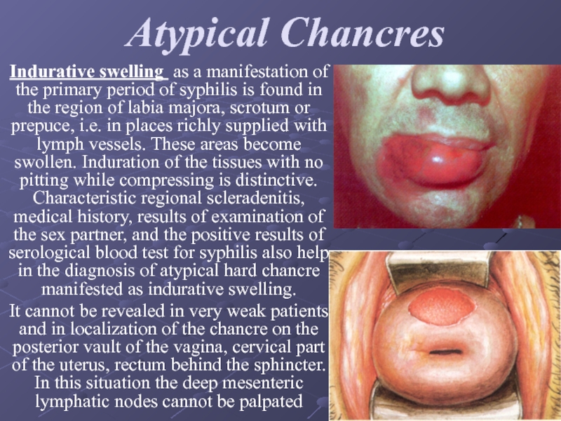
Indurative swelling as a manifestation of the primary period of
syphilis is found in the region of labia majora, scrotum or prepuce, i.e. in places richly supplied with lymph vessels. These areas become swollen. Induration of the tissues with no pitting while compressing is distinctive. Characteristic regional scleradenitis, medical history, results of examination of the sex partner, and the positive results of serological blood test for syphilis also help in the diagnosis of atypical hard chancre manifested as indurative swelling.
It cannot be revealed in very weak patients and in localization of the chancre on the posterior vault of the vagina, cervical part of the uterus, rectum behind the sphincter. In this situation the deep mesenteric lymphatic nodes cannot be palpated
It cannot be revealed in very weak patients and in localization of the chancre on the posterior vault of the vagina, cervical part of the uterus, rectum behind the sphincter. In this situation the deep mesenteric lymphatic nodes cannot be palpated
Слайд 10Complications of hard chancre
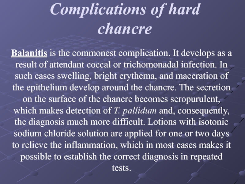
Balanitis is the commonest complication. It develops as
a result of attendant coccal or trichomonadal infection. In such cases swelling, bright erythema, and maceration of the epithelium develop around the chancre. The secretion on the surface of the chancre becomes seropurulent, which makes detection of T. pallidum and, consequently, the diagnosis much more difficult. Lotions with isotonic sodium chloride solution are applied for one or two days to relieve the inflammation, which in most cases makes it possible to establish the correct diagnosis in repeated tests.
Слайд 11Complications of hard chancre
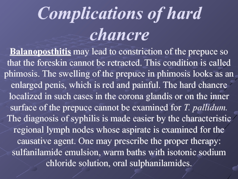
Balanoposthitis may lead to constriction of the prepuce
so that the foreskin cannot be retracted. This condition is called phimosis. The swelling of the prepuce in phimosis looks as an enlarged penis, which is red and painful. The hard chancre localized in such cases in the corona glandis or on the inner surface of the prepuce cannot be examined for T. pallidum. The diagnosis of syphilis is made easier by the characteristic regional lymph nodes whose aspirate is examined for the causative agent. One may prescribe the proper therapy: sulfanilamide emulsion, warm baths with isotonic sodium chloride solution, oral sulphanilamides.
Слайд 12Complications of hard chancre
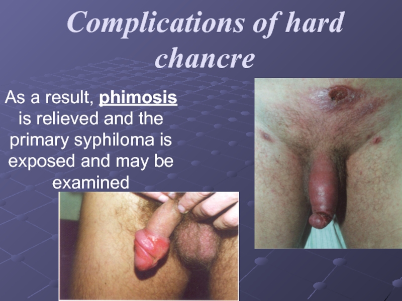
As a result, phimosis is relieved and the
primary syphiloma is exposed and may be examined
Слайд 13Complications of hard chancre
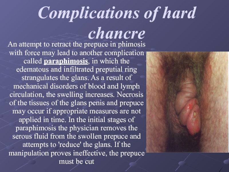
An attempt to retract the prepuce in phimosis
with force may lead to another complication called paraphimosis, in which the edematous and infiltrated preputial ring strangulates the glans. As a result of mechanical disorders of blood and lymph circulation, the swelling increases. Necrosis of the tissues of the glans penis and prepuce may occur if appropriate measures are not applied in time. In the initial stages of paraphimosis the physician removes the serous fluid from the swollen prepuce and attempts to 'reduce' the glans. If the manipulation proves ineffective, the prepuce must be cut
Слайд 14Complications of hard chancre
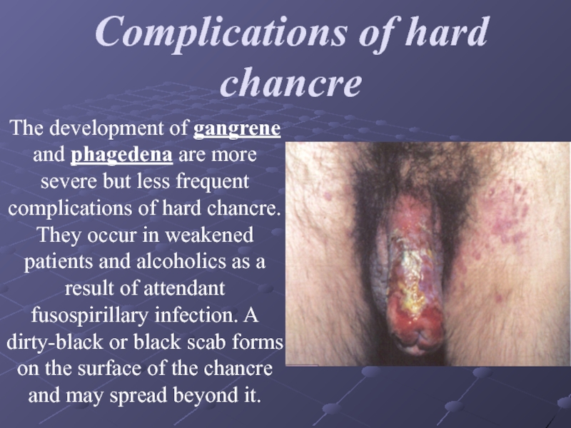
The development of gangrene and phagedena are more
severe but less frequent complications of hard chancre. They occur in weakened patients and alcoholics as a result of attendant fusospirillary infection. A dirty-black or black scab forms on the surface of the chancre and may spread beyond it.
Слайд 15Complications of hard chancre
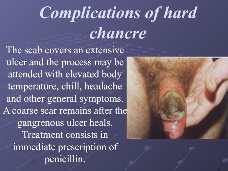
The scab covers an extensive ulcer and the
process may be attended with elevated body temperature, chill, headache and other general symptoms. A coarse scar remains after the gangrenous ulcer heals. Treatment consists in immediate prescription of penicillin.
Слайд 16Secondary syphilis
Secondary syphilis develops 2.5-3, rarely 4 months later after infection.
Without treatment relapses can occur during 2-4 years and longer (the first relapse most often develops 4-6 months after infection).
In practical venereology there are situations when the T. pallidum enters the blood directly (transfusion from the donor with syphilis). In such patients after 2-2.5 hours the lesions characteristic of secondary syphilis, with preliminary prodromal occurrences, are formed on the skin and mucous membranes.
In practical venereology there are situations when the T. pallidum enters the blood directly (transfusion from the donor with syphilis). In such patients after 2-2.5 hours the lesions characteristic of secondary syphilis, with preliminary prodromal occurrences, are formed on the skin and mucous membranes.
Слайд 17Secondary Siphilis
Characteristic signs of secondary syphilis
Focal situation of the lesions.
Round form
and sharp borders of the lesions.
Specific colour (“ham coloured” or “copper-red”).
Lesions are different in morphology (polymorphism)
Absence of subjective feelings.
Progress of the lesions without fever.
Tendency towards voluntary reverse development.
Soon cured by specific therapy.
Specific colour (“ham coloured” or “copper-red”).
Lesions are different in morphology (polymorphism)
Absence of subjective feelings.
Progress of the lesions without fever.
Tendency towards voluntary reverse development.
Soon cured by specific therapy.
Слайд 18Secondary syphilis
Macular syphilis, or syphilitic roseola. Clinical and morphological characteristics of
syphilitic roseolaThe following clinical forms of roseola should be differentiated:
a) small macular,
b) large macular,
c) nettle rash,
d) spotted or granular,
e) fused,
f) circle-shaped, or ring-shaped.
a) small macular,
b) large macular,
c) nettle rash,
d) spotted or granular,
e) fused,
f) circle-shaped, or ring-shaped.
Слайд 19
Diagnosis of macular syphilid (roseola) is based on its characteristic features:
vascular macula, which disappears by pressing, and, as a rule, does not rise above the level of the skin
pinkish red colour with the blue font;
size of the nail of the little finger;
roseola is situated on the sides of the trunk, chest, abdomen, upper limbs (roseola does not develop on the skin of the face, feet, hands);
roseola develops slowly and disappears after 1-2 weeks. In some cases roseola can rise above the level of the skin slowly or temporarily. In urticarial roseola the patients may feel itch, burning sensation;
during the regress of roseola there may be squamation. Granular roseola and hemorrhagic roseola develop in patients with high penetration of blood vessels.
Слайд 20Papular syphilid
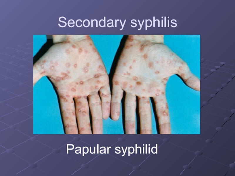
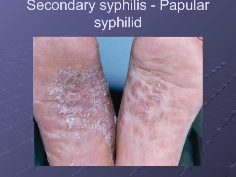
is often a sign of secondary, usually relapsing syphilis.
The papule is situated in the stratum papillaris of the dermis.
The characteristic features of the papules are hemispherical form, sharp borders, even contours, ham colour; they do not fuse with each other.
Слайд 22
The following clinical varieties of papular lesions are differentiated:
a) miliary
B) lenticular
C)
coin-shaped
D) condyloma latum
E) psoriatic form
The papules can be situated anywhere (skin, mucous membranes, rarely on the vocal ligaments: syphilitic hoarse throat).
D) condyloma latum
E) psoriatic form
The papules can be situated anywhere (skin, mucous membranes, rarely on the vocal ligaments: syphilitic hoarse throat).
Слайд 24
Pustular syphilid.
Pustular syphilid occurs rarely and develops in weak patients and
in alcoholics. The following types of pustular syphilid are differentiated:
1) syphilitic impetigo
2) varioliform syphilid
3) syphilitic ecthyma
4) syphilitic rupia
5) acneform
1) syphilitic impetigo
2) varioliform syphilid
3) syphilitic ecthyma
4) syphilitic rupia
5) acneform
Слайд 25
Syphilitic alopecia or calvities
Alopecia areata: diffuse and mixed forms are differentiated.
Syphilitic alopecia can be observed in 15-18% of patients with secondary or relapsing syphilis. In alopecia areata on the head small, rounded contoured foci of baldness without signs of inflammation and squamation are observed. In diffused baldness the hair fall out equally from all parts of the head. The Pinkus’ sign: syphilitic alopecia in the region of beard, eyebrows, eye lashes.
Слайд 26Syphilitic leukoderma, or pigmentary syphilid (leukoderma syphiliticum)
Pigmentary syphilid appears in patients
not earlier than 5 to 6 months after infection, i.e. in the secondary recurrent period. Whitish, as if depigmented, round or oval spots resembling lace-work or a net, are formed on hyperpigmented skin on the sides and back of the neck, in the axillae, and on the sides of the chest. As a rule, abnormalities are found in the cerebrospinal fluid, but the available methods of examination yield no convincing data on clinical affection of the nervous system of most patients with syphilitic leukoderma.

