- Главная
- Разное
- Дизайн
- Бизнес и предпринимательство
- Аналитика
- Образование
- Развлечения
- Красота и здоровье
- Финансы
- Государство
- Путешествия
- Спорт
- Недвижимость
- Армия
- Графика
- Культурология
- Еда и кулинария
- Лингвистика
- Английский язык
- Астрономия
- Алгебра
- Биология
- География
- Детские презентации
- Информатика
- История
- Литература
- Маркетинг
- Математика
- Медицина
- Менеджмент
- Музыка
- МХК
- Немецкий язык
- ОБЖ
- Обществознание
- Окружающий мир
- Педагогика
- Русский язык
- Технология
- Физика
- Философия
- Химия
- Шаблоны, картинки для презентаций
- Экология
- Экономика
- Юриспруденция
Face department of the head part 1 презентация
Содержание
- 1. Face department of the head part 1
- 2. face skeleton The volume of an obverse
- 3. Face Fascies and Cellulose space In face
- 4. Face Fascies Superficial fascia persons the thin
- 5. fascia parotideomasseterica In area parotis salivary gland
- 6. Face Fascies In area parotis salivary gland
- 7. borders of Parotideomasseteric region Upper-zygomatic arch In
- 9. In parotid gland are located External carotid
- 10. Parotideomasseteric region
- 11. weak places of a capsule of parotid
- 12. Transverse cut through parotid gland. (Internal pharyngeal part)
- 13. Your conclusions about inflammatory process in parotid gland
- 14. Inflammatory process in parotid gland Local (fascial
- 15. Face department of the head part 2
- 16. Borders of buccal areas Above – Margo
- 17. Superficial fascia Covers mimic muscles Forms a
- 18. Fascia proper In front from a
- 19. Corpus adipose buccal It is well expressed
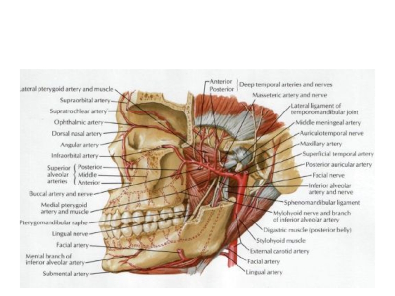
- 20. Superficial vessels buccal areas Facial artery and
- 21. Facial artery and vien
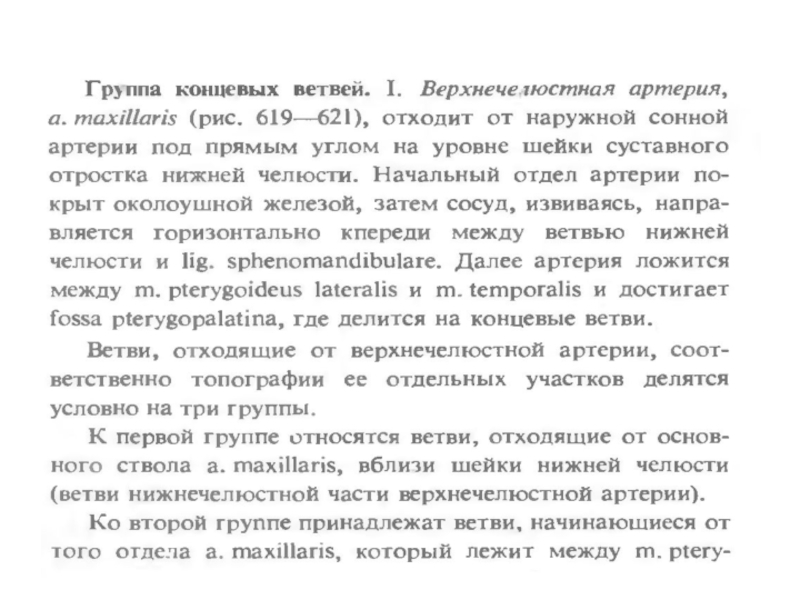
- 22. Face department of the head part 3
- 23. Фасции лица Глубокий листок собственной фасции лица
- 24. Нижнечелюстной нерв и верхнечелюстная артерия
- 25. Глубокая область лица Занимает подвисочную ямку.
- 26. Содержит Латеральную и медиальную крыловидные мышцы и
- 27. жевательные мышцы
- 28. Глубокие жевательные мышцы
- 29. Артерия maxillaris
- 30. Вены лица
- 31. Сообщения крылонебной ямки С ротовая полостью –через
- 32. Клетчаточные пространства глотки В окологлоточном пространстве различают
- 33. Сосуды и нервы глубокой области лица
- 34. Сосуды и нервы
- 44. Связи венозной системы лица и синусов тв. мозг. оболочки
- 45. Мимические мышцы
- 46. Мышцы ротовой щели
- 47. Ветви лицевого нерва Височная Скуловая Щечная Краевая нижней челюсти Шейная Задняя ушная
- 48. Мимические мышцы и лицевой нерв
- 49. occipitofrontalis
- 50. Orbicularis oculi
- 51. Buccinator +orbicularis oris
- 52. Zigomaticus major + minor
- 53. Depressor labii+levator labii superior
- 54. Rizorius (смеха)
- 55. Зоны иннервации тройничного нерва
- 56. Тройничный нерв и шейное сплетение
- 57. сосуды
- 58. вены
- 59. Мышцы глазного яблока
- 60. Мышцы глазного яблока
- 61. Глубокая область лица
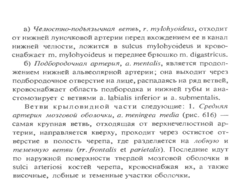
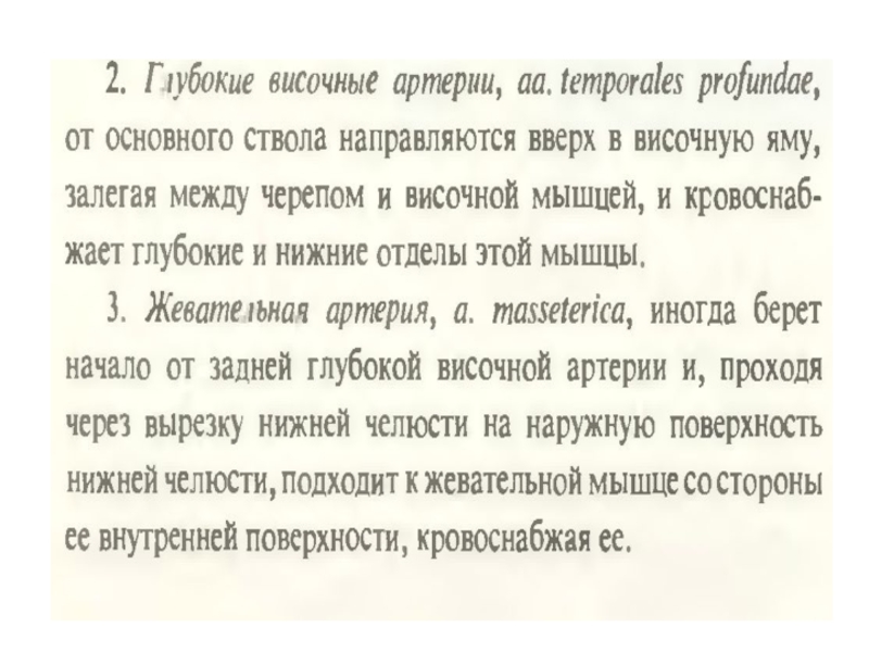
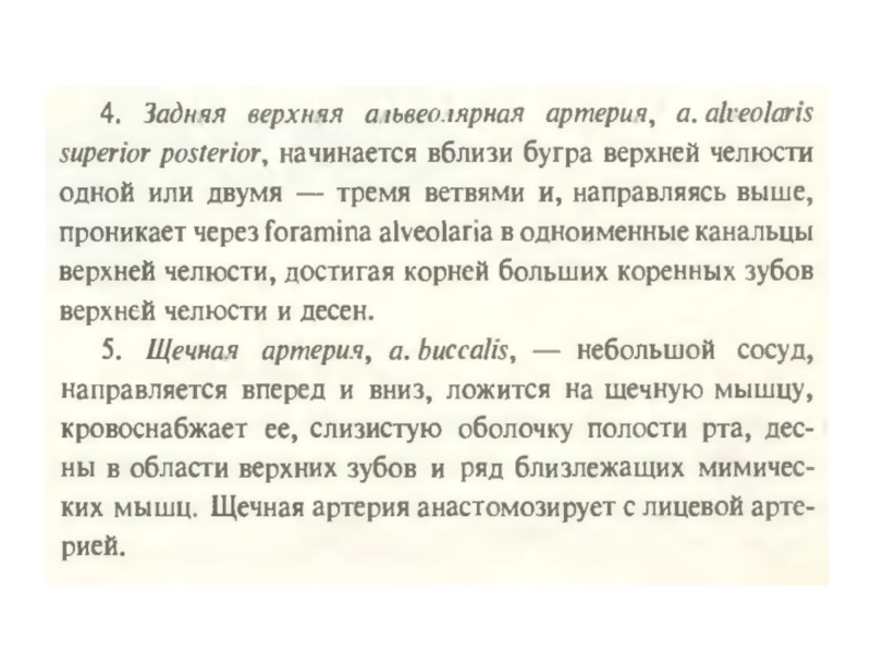
- 62. Ветви артерии maxillaris
- 63. n.Maxillaris и mandibularis
- 64. Проекция ветвей тройничного нерва
- 67. Лимфатическая система лица и шеи
- 68. Твердое и мягкое небо
- 69. Мягкое небо
- 70. Мышцы мягкого неба
- 71. Латеральная стенка полости носа
- 72. раковины
- 73. Придаточные пазухи
- 74. Придаточные пазухи, вид со стороны глазницы
- 75. Медиальная стенка полости носа (перегородка)
- 76. Иннервация латеральной стенки носа
- 77. Иннервация медиальной стенки носа (перегородка)

Слайд 2face skeleton
The volume of an obverse skeleton at children about one
year makes – 13 % of volume of a cranium, at adults – 40 %.
In extreme old age the skull loses to 40 % of the weight. Growth of obverse bones and definitive formation of outlines of the person comes to the end by 20-23 years at men, by 16-18 – at women.
Thickness of a skin – from 0,5 to 4 mm. The skin of newborns in 3,5 times is more thin a skin of adults. The skin is similar to a face skin retroauricular areas, necks, over - and subclavicular areas, an internal surface of a shoulder, a lateral surface of a thorax which use for plastic operations on the person.

Слайд 3Face Fascies and Cellulose space
In face department of a head allocate
fascia which limit cellular spaces. Except basic function fascia matter in distribution of inflammatory processes and hematomas
Fatty cellular it is favorable for development of a purulent inflammation. If we know fascies we can understand dynamics and a direction of inflammatory process.
Especially it is a lot of fascies and fatty congestions is available in the field of an eye-socket, a mouth floor, lateral departments of the face and around pharynx
Face department of the head Face department of the head

Слайд 4Face Fascies
Superficial fascia persons the thin and forms cases for mimic
muscles and neurovascular formations.
Hypodermic cellular it is located between a skin and superficial fascia, at different people its thickness is various, but it is especially developed at children.
Between separate mimic muscles there is variously expressed layer fatty cellular.
Proper fascia lateral area of the person divided on superficial and deep.

Слайд 5fascia parotideomasseterica
In area parotis salivary gland the superficial lamina proper fascie
forms its capsule and carries the name fascia parotidea, It is continuation temporal fascia.
In front from parotis glands this fascia covers a chewing muscle and carries the name chewing fascia {fascia masseterica).
Together they form fascia parotideomasseterica.
At back edge parotis glands fascia it is fixed to a cartilage of external acoustical porus and to an external surface of a corner of the mandible, then passes on mastoid processes.

Слайд 6Face Fascies
In area parotis salivary gland from fascia depart into glands
septum. They divide it into separate parts.
Fascia covers a chewing muscle and also gives to it partitions, but in some places, especially on an internal surface of a muscle fascia easily departs from the mandible bon.
Here it is located the space. Its name masseterico-mandibular
Below this space is closed by fixing of a masseter muscle to mandibul bones, and up passes in space of temporal area.

Слайд 7borders of Parotideomasseteric region
Upper-zygomatic arch
In front-anterior part of masseter muscles
Behind-vertical line
through external acoustical porus
From below - along Margo mandible to a Bone corner and then to top of mastoid process
From below - along Margo mandible to a Bone corner and then to top of mastoid process

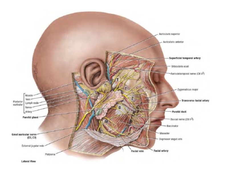
Слайд 9In parotid gland are located
External carotid artery
Maxillary artery
Superficial temporal artery and
there branch – transversal facial artery
Facial nerve
Auricle-temporal nerve
Lymph nodes
Facial nerve
Auricle-temporal nerve
Lymph nodes


Слайд 11weak places of a capsule of parotid gland
Internal surface (there is
not capsule)
The top site which lies near to external acoustical pass
The top site which lies near to external acoustical pass
The top site")
")

Слайд 14Inflammatory process in parotid gland
Local (fascial septum)
When a patient have purulent
parotitis - In 2 case from 3 pus extends on pharyngeal part of parotid gland and then to mediastinum (53% patients die)
In 1 case from 3 pus extends to porus acusticus externus (follows outside)
Damage of vessels and nerves is possible. (which lie inside)
In 1 case from 3 pus extends to porus acusticus externus (follows outside)
Damage of vessels and nerves is possible. (which lie inside)
When a patient have purulent parotitis - In 2")

Слайд 16Borders of buccal areas
Above – Margo inferior orbital
Below – Margo inferior
mandibule
In front – Plica naso-buccalis and naso-labialis
Behind – margo anterior masseter
In front – Plica naso-buccalis and naso-labialis
Behind – margo anterior masseter

Слайд 17Superficial fascia
Covers mimic muscles
Forms a case for a lump of Bisha
(being split on 2 leaves)
")
Слайд 18Fascia proper
In front from a masseter muscle fascia proper covered buccal
muscle. This part carries the name fascia buccopharyngea
Behind- the deep part of buccal fascia covers to a lateral surface of faring.
Behind- the deep part of buccal fascia covers to a lateral surface of faring.

Слайд 19Corpus adipose buccal
It is well expressed at small children. A capsule
for it forms superficial fascia.
There are 3 processes at a lump of Bisha. On these ways the infection can get to deep layers of a head and the face.
Shoots of corpus adipose buccal (Bisha)
Temporal (Proceeds to subaponeurotic space temporal region)
Ophtalmic ( to inferior orbital fissure and to orbita)
Pterygopalatine (to pterygopalatine fossa, then 5 ways : 1-orbita, 2-nasal cavity, 3-oral cavity, 4-internal base of skull, 5- external base of skull )

Слайд 20Superficial vessels buccal areas
Facial artery and vеin.
Artery has many bends
Vein
– forward stroke
Vienna – a straight line
Projection – between Margo anterior masseter muscle and an internal corner of an eye
Vienna – a straight line
Projection – between Margo anterior masseter muscle and an internal corner of an eye


Слайд 23Фасции лица
Глубокий листок собственной фасции лица носит название межкрыловидной фасции. Она
отделяет друг от друга крыловидные мышцы и имеет направление сверху вниз, изнутри кнаружи, спереди назад, таким образом располагаясь в косой плоскости. Эта фасция начинается от крыловидного отростка основной кости, вдоль внутреннего края овального и остистого отверстия до угловой ости. Прикрепляется эта фасция в месте прикрепления медиальной крыловидной мышцы. Утолщения межкрыловидной фасции образуют связки — крыло-челюстную, крыло-основную и основно-челюстную.


Слайд 25Глубокая область лица
Занимает подвисочную ямку.
Ограничена:
Вверху-большое крыло клиновидной кости
Спереди-бугор верхней челюсти
Сзади-околоушная
железа
Снизу-ограничена прикреплением к нижней челюсти медиальной крыловидной мышцы
Снизу-ограничена прикреплением к нижней челюсти медиальной крыловидной мышцы

Слайд 26Содержит
Латеральную и медиальную крыловидные мышцы и конечный (нижний) участок височной мышцы
Верхнечелюстную
артерию
Крыловидное венозное сплетение
3 пространства-височно-крыловидное с артерией и венозным сплетением,межкрыловидное с нижнечелюстным нервом и крылонебная ямка с верхнечелюстным нервом
Крыловидное венозное сплетение
3 пространства-височно-крыловидное с артерией и венозным сплетением,межкрыловидное с нижнечелюстным нервом и крылонебная ямка с верхнечелюстным нервом
 участок височной мышцыВерхнечелюстную артериюКрыловидное венозное сплетение3 пространства-височно-крыловидное")


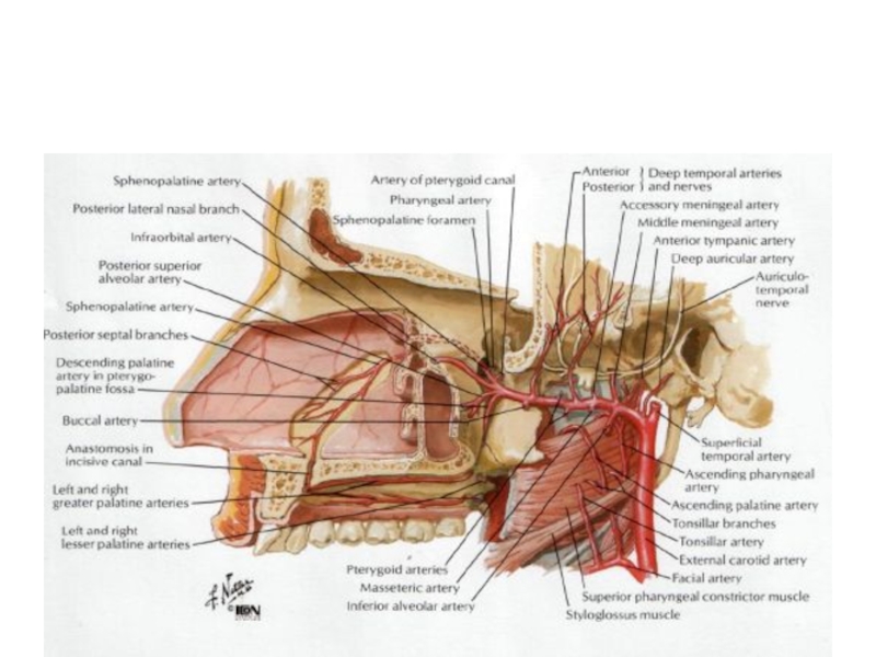
Слайд 31Сообщения крылонебной ямки
С ротовая полостью –через большой небный канал
С полостью глазницы
-через нижнюю глазничную щель
С полостью носа – через foramen sphenopalatina (одноименная артерия)
Со средней черепной ямкой – через круглое отверстие (2-я ветвь тройничного нерва)
С наружным основанием черепа –через крыловидный канал
С полостью носа – через foramen sphenopalatina (одноименная артерия)
Со средней черепной ямкой – через круглое отверстие (2-я ветвь тройничного нерва)
С наружным основанием черепа –через крыловидный канал

Слайд 32Клетчаточные пространства глотки
В окологлоточном пространстве различают передний и задний отделы. Границу
между ними образует шиловидный отросток с начинающимися от него шиловидно-глоточной, шиловидно-язычной и шиловидно-подъязычной мышцами и фасциальный листок, натянутый между шиловидным отростком и глоткой (шиловидно-глоточный апоневроз).
К переднему отделу парафаренгиального пространства примыкают: снутри – небная миндалина, снаружи – глоточный отросток околоушной железы. В этом пространстве располагаются ветви восходящей небной артерии и одноименные вены, играющие роль в распространении воспалительного процесса из района миндалин (при перитонзиллярном абсцессе).
В заднем отделе парафаренгиального пространства проходят внутренняя яремная вена, внутренняя сонная артерия, IX, X, XI, XII черепные нервы, симпатический ствол и верхние глубокие шейные лимфатические узлы.
Заглоточное пространство делится по середине перегородкой на правый и левый отделы, поэтому заглоточные абсцессы бывают обычно односторонними.


")


")

")