- Главная
- Разное
- Дизайн
- Бизнес и предпринимательство
- Аналитика
- Образование
- Развлечения
- Красота и здоровье
- Финансы
- Государство
- Путешествия
- Спорт
- Недвижимость
- Армия
- Графика
- Культурология
- Еда и кулинария
- Лингвистика
- Английский язык
- Астрономия
- Алгебра
- Биология
- География
- Детские презентации
- Информатика
- История
- Литература
- Маркетинг
- Математика
- Медицина
- Менеджмент
- Музыка
- МХК
- Немецкий язык
- ОБЖ
- Обществознание
- Окружающий мир
- Педагогика
- Русский язык
- Технология
- Физика
- Философия
- Химия
- Шаблоны, картинки для презентаций
- Экология
- Экономика
- Юриспруденция
Esophageal Cancer презентация
Содержание
- 1. Esophageal Cancer
- 2. Esophageal Cancer Epidemiology and Risk
- 3. Epidemiology Over 15,000 patients per
- 4. Incidence of Esophageal Cancer
- 5. Adenocarcinoma: Barrett’s Esophagus Likely related
- 6. Barrett’s Esophagus and Esophageal Cancer
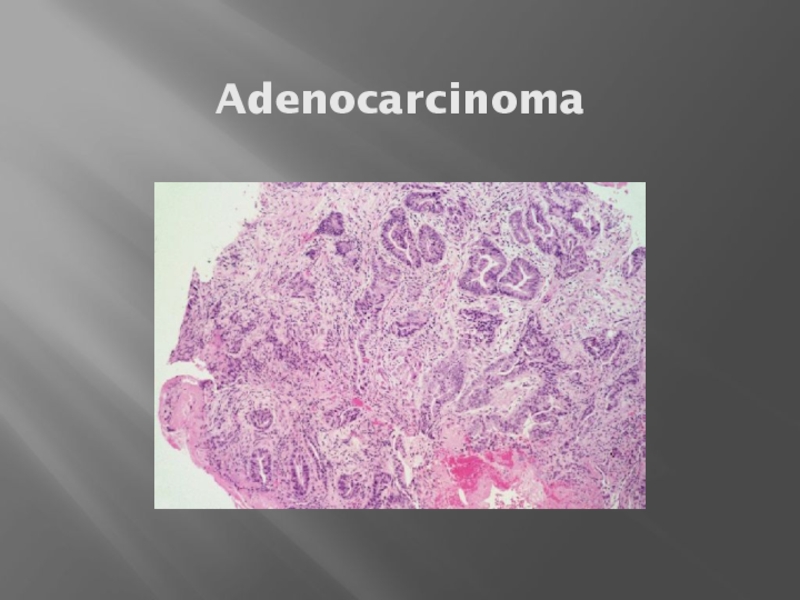
- 7. Adenocarcinoma
- 9. Squamous Cell Carcinoma Usually upper
- 10. Squamous Cell Carcinoma
- 11. Anatomy
- 12. Clinical Presentation Signs: weight loss,
- 13. Endoscopy ENDOSCOPIC IMAGE OF
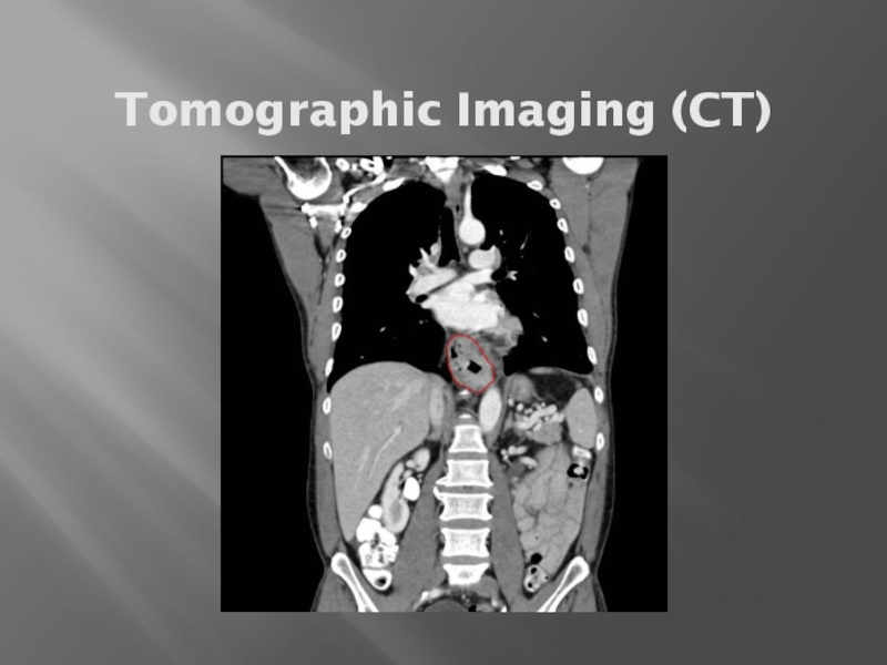
- 14. Tomographic Imaging (CT)
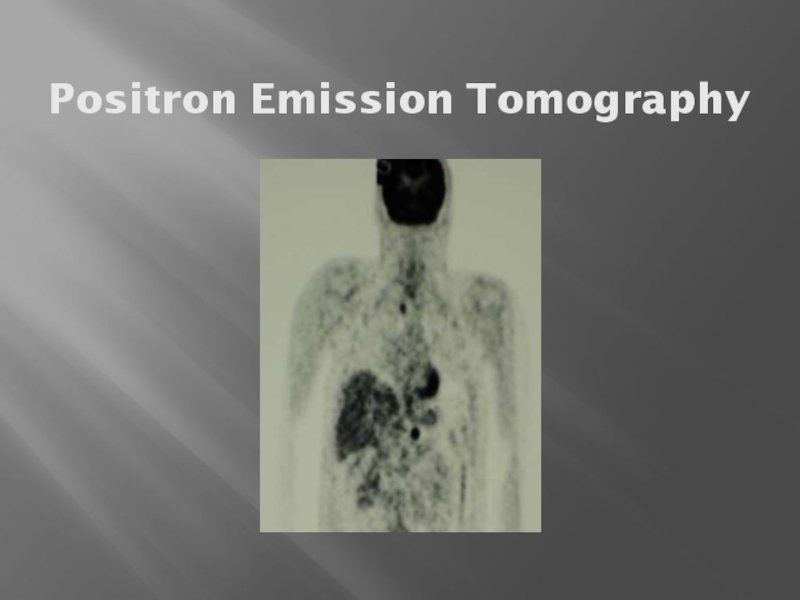
- 15. Positron Emission Tomography
- 16. Staging Two basic groups Locally
- 17. Locally Advanced Stage “Best” treatment
- 18. Chemotherapy & Radiation Without Surgery 5y
- 19. Pattern of Recurrence Almost always
- 20. Treatment of Metastatic Disease Palliative
- 21. Palliation For swallowing trouble: stent
- 22. Molecular Markers/Targets
Слайд 2
Esophageal Cancer
Epidemiology and Risk Factors
Diagnosis — signs, symptoms, and tests
Work-up
Treatment Overview
Future
Directions
Слайд 3
Epidemiology
Over 15,000 patients per year in the United States and 7th
leading cause of cancer death in men.
8th most common cancer worldwide.
Most cases are squamous cell, related to tobacco and alcohol exposure.
In Western countries, adenocarcinoma increasing thought due to Barrett’s esophagus.
Approximately 50% present with advanced disease, which is incurable.
8th most common cancer worldwide.
Most cases are squamous cell, related to tobacco and alcohol exposure.
In Western countries, adenocarcinoma increasing thought due to Barrett’s esophagus.
Approximately 50% present with advanced disease, which is incurable.
Слайд 5
Adenocarcinoma: Barrett’s Esophagus
Likely related to chronic GERD, obesity.
Pathway of malignant progression.
40
to 125 times relative risk of adenocarcinoma.
Incidence of cancer is approximately 0.5% per year in patients with BE.
No known effective screening tool.
Usually Lower esophagus/GE junction.
Incidence of cancer is approximately 0.5% per year in patients with BE.
No known effective screening tool.
Usually Lower esophagus/GE junction.
Слайд 6
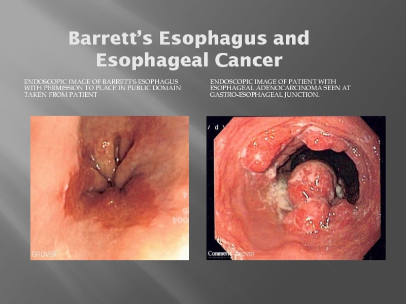
Barrett’s Esophagus and Esophageal Cancer
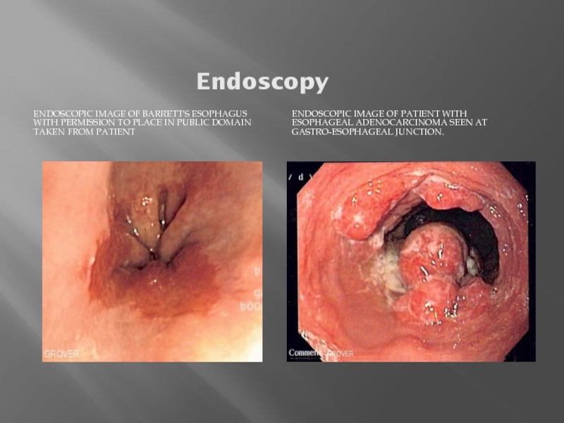
ENDOSCOPIC IMAGE OF BARRETT'S ESOPHAGUS WITH PERMISSION
TO PLACE IN PUBLIC DOMAIN TAKEN FROM PATIENT
ENDOSCOPIC IMAGE OF PATIENT WITH ESOPHAGEAL ADENOCARCINOMA SEEN AT GASTRO-ESOPHAGEAL JUNCTION.
Слайд 9
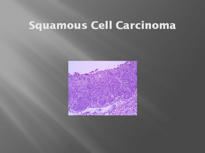
Squamous Cell Carcinoma
Usually upper and middle esophagus.
Tends to be a local
problem—less metastases.
Most common worldwide histology.
Carcinogens present in tobacco and alcohol.
Most common worldwide histology.
Carcinogens present in tobacco and alcohol.
Слайд 12
Clinical Presentation
Signs: weight loss, palpable lymph nodes, usually non-specific.
Symptoms: dysphagia, loss
of appetite, pain with swallowing, fatigue, cough, retrosternal and abdominal pain.
Lab Data: no tumor markers.
Lab Data: no tumor markers.
Слайд 13
Endoscopy
ENDOSCOPIC IMAGE OF BARRETT'S ESOPHAGUS WITH PERMISSION TO PLACE IN PUBLIC
DOMAIN TAKEN FROM PATIENT
ENDOSCOPIC IMAGE OF PATIENT WITH ESOPHAGEAL ADENOCARCINOMA SEEN AT GASTRO-ESOPHAGEAL JUNCTION.
Слайд 16
Staging
Two basic groups
Locally Advanced (primary tumor and regional lymph nodes):
- potentially curable
Metastatic (distant spread)
-Incurable
-survival increased with chemotherapy
Metastatic (distant spread)
-Incurable
-survival increased with chemotherapy
Слайд 17
Locally Advanced Stage
“Best” treatment approach is controversial and continually evolving.
Concepts to
consider:
Local control (primary tumor)
Distant disease (“micrometastases”)
Modes of treatment include surgery, radiation and chemotherapy in various sequences and combinations
Local control (primary tumor)
Distant disease (“micrometastases”)
Modes of treatment include surgery, radiation and chemotherapy in various sequences and combinations
Слайд 18
Chemotherapy & Radiation Without Surgery
5y survival:
radiation therapy only - 0%
Combination
treatment – 26%
Survival and Pathologic Response
Survival and Pathologic Response
Слайд 19
Pattern of Recurrence
Almost always at a distant site.
Approaches to this problem.
Adjuvant chemotherapy
Newer chemotherapy
Induction chemotherapy
Intensified chemotherapy
Result: nothing is much better…
Newer chemotherapy
Induction chemotherapy
Intensified chemotherapy
Result: nothing is much better…
Слайд 20
Treatment of Metastatic Disease
Palliative
No standard chemotherapy approach
Combination of two drugs based
on 5-FU, platins, taxanes.
-Cisplatin/CPT-11, FOLFOX
Median survival ~ 9 months
Clinical trial
-Cisplatin/CPT-11, FOLFOX
Median survival ~ 9 months
Clinical trial
Слайд 21
Palliation
For swallowing trouble: stent most common
For pain: narcotics, radiation
For Cachexia: appetite
stimulants, feeding tubes

