- Главная
- Разное
- Дизайн
- Бизнес и предпринимательство
- Аналитика
- Образование
- Развлечения
- Красота и здоровье
- Финансы
- Государство
- Путешествия
- Спорт
- Недвижимость
- Армия
- Графика
- Культурология
- Еда и кулинария
- Лингвистика
- Английский язык
- Астрономия
- Алгебра
- Биология
- География
- Детские презентации
- Информатика
- История
- Литература
- Маркетинг
- Математика
- Медицина
- Менеджмент
- Музыка
- МХК
- Немецкий язык
- ОБЖ
- Обществознание
- Окружающий мир
- Педагогика
- Русский язык
- Технология
- Физика
- Философия
- Химия
- Шаблоны, картинки для презентаций
- Экология
- Экономика
- Юриспруденция
Disorders of Calcium Metabolism презентация
Содержание
- 1. Disorders of Calcium Metabolism
- 2. Calcium An essential intracellular and extracellular cation
- 3. calcium 40-45 % ionized 40-45
- 4. Calcium and Albumin 40-45% of circulating
- 5. Calcium and Albumin At pH 7.4 each
- 6. Disorders of Calcium Metabolism Hypercalcemia Serum
- 7. Causes of hypercalcemia PTH-mediated Primary
- 8. Causes of hypercalcemia Vitamin D intoxication
- 9. Diagnosis of Hypercalcemia Serum Ca ↑
- 10. Manifestations of hypercalcemia
- 11. Renal manifestations The most important renal manifestations
- 12. Renal manifestations Nephrolithiasis — When hypercalcemia is
- 13. Gastrointestinal manifestations Nausea. Constipation may
- 14. Cardiovascular symptoms Shortened QT interval Ventricular
- 15. Hyperparathyroidism Secondary hyperparatroidism: due to vitamin
- 16. Primary Hyperparathyroidism Incidence 1/1000- 42/100 000 Postmenopausal
- 17. or multiple adenomas, nearly 100% 15-20% up to 60% nearly 100% 30-100% 10-25%
- 18. Clinical Manifestations of Prim Hyperparathyroidism CNS -
- 19. Bone and Joint Manifestations in Primary Hyperparathyroidism
- 22. Treatment When is surgery indicated in PHPT patients ?
- 23. Guidelines for Surgery
- 24. Other Considerations Neuropsychological disturbances Weakness and easy
- 25. US in the diagnosis of PTA
- 26. 99mTc- sestamibi Parathyroid Scan 20
- 27. What is 4D-CT? 4D-CT includes image
- 28. 4D CT in diagnosis of PHPT Arterial
- 29. Patients who weren‘t operated- Monitoring guidelines
- 30. General Measures Hydration Adequate Mobility Diet neither
- 31. Calcium-sensing Receptor A member of the G
- 32. Inactivating Mutations in Calcium Sensing Receptor Inactivating
- 33. Hypercalcemia of Malignancy Lung, breast, and prostate
- 34. PthrP Induced Hypercalcemia Squamous cell carcinoma Islet
- 35. PTHrP-PTH related peptide Gen located on the
- 36. Treatment Hydration Bisphosphonates IV, Denosumab Calcitonin Glucocorticoids Dialysis
- 37. Hydration First step in the management of
- 38. Loop Diuretics In the past intensive administration
- 39. Calcitonin The efficacy of calcitonin (4 IU/kg
- 40. Bisphosphonates Structurally related to pyrophosphate Great affinity
- 41. Zoledronic Acid (ZOMERA) Zoledronic acid belongs to
- 42. Denosumab Mechanism of Action © 2007 Amgen.
- 43. Inhibit the growth of neoplastic lymphoid tissue Counteract the effects of vitamin D Glucocorticoids
- 44. Low PTH Hypocalcemia
- 45. Hypocalcemia Low PTH (hypoparathyroidism) Genetic disorders:
- 46. Hypocalcemia High PTH (secondary hyperparathyroidism in response
- 47. Drugs induced hypocalcemia Inhibitors of bone resorption
- 48. Trousseau's sign is the induction of carpopedal
- 49. Hypocalcemia: Carpopedal spasm and ECG changes Carpopedal
- 50. Treatment of Acute Hypocalcemia Promptly correct symptomatic
- 51. Treatment of Acute Hypocalcemia For those with
- 52. To prevent Hypocalcemia due to Hungry bone
- 53. Primary hypoparathyroidism
- 58. Natpara Natpara- for use in patients who
- 59. Causes of magnesium depletion Causes of magnesium depletion Renal losses Gasro-intestinal losses
- 60. Treatment of Hypomagnesemia If the serum
- 61. Treatment of Hypomagnesemia Oral repletion- if
- 62. Thank you and Good luck!

Слайд 2Calcium
An essential intracellular and extracellular cation
Extracellular calcium is required to maintain
Intracellular calcium is needed for normal activity of many enzymes.
Regulation of endocrine and exocrine secretory activities: insulin, aldosterone
Activation of compliment system
Bone metabolism: calcium salts provide structural integrity of the skeleton ( mineralization)

Слайд 3
calcium
40-45 %
ionized
40-45 %
albumin
bound
10%
complexed
citrate, sulphate
8.5–10.5 mg/dl
ECF
Filtration
5-7gr
Reabsorption
4.9-6.7gr
98%
Diet 0.5-1.5 gr
Absorption
0.25-0.5gr
Secretion
0.1-0.2gr
Feces
0.35-0.6gr
Resorption
0.3-0.5 g
Formation
0.3-0.5 g
Bone
1000
0.15-0.3 g/24h
Total body Ca
1 to 1.5 kg
99%- skeleton
0.1% ECF
rest intracellular

Слайд 4Calcium and Albumin
40-45% of circulating calcium is bound to albumin
Change
Calcium is bound to carboxyl groups in albumin, this binding is highly pH dependent
Acute acidosis binding ionized calcium
Acute alkalosis binding ionized calcium
A shift of 0.1 pH unit produces a change in ionized calcium of 0.04 to 0.05 mmol/L ( 0.16-0.2 mg/dl)
These changes are not reflected at the total calcium

Слайд 5Calcium and Albumin
At pH 7.4 each g/dl of serum albumin binds
Serum calcium should be “corrected” according to serum albumin level
Normal level of serum albumin is 4 g/dl
Change of 1 g/dl in serum albumin 0.8 mg/dl in total serum calcium
Example: A patient with total serum calcium 7.5 mg/dl albumin 2 g/dl, has corrected serum calcium of 9.1 mg/dl
Ca = SerumCa + 0.8 * (NormalAlbumin - PatientAlbumin)

Слайд 6Disorders of Calcium Metabolism
Hypercalcemia Serum Calcium >10.4 mg/dl
Hypocalcemia

Слайд 7Causes of hypercalcemia
PTH-mediated
Primary hyperparathyroidism (sporadic) –solitary adenoma or hyperplasia
Familial MEN1
FHH
Tertiary hyperparathyroidism (renal failure)
PTH-independent
Hypercalcemia of malignancy:
PTHrp
Activation of extrarenal 1 alpha-hydroxylase (increased calcitriol)
Osteolytic bone metastases and local cytokines
 –solitary adenoma or hyperplasiaFamilial MEN1 and -2a FHH")
Слайд 8Causes of hypercalcemia
Vitamin D intoxication
Chronic granulomatous disorders: Activation of extrarenal 1
Medications :
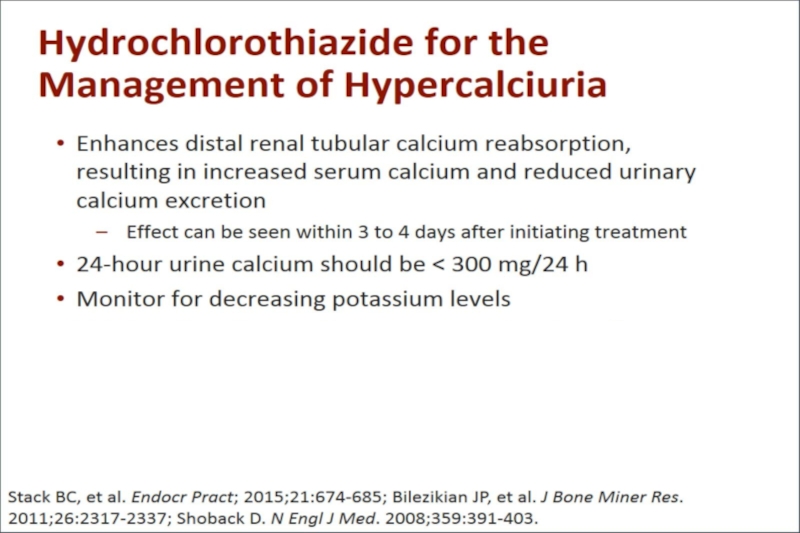
Thiazide diuretics
Lithium
Teriparatide
Excessive vitamin A
Theophylline toxicity
Associated with high bone turnover
Hyperthyroidism
Immobilization
Acromegaly
Miscellaneous Pheochromocytoma Adrenal insufficiency Parenteral nutrition
Milk alkali syndrome
")
Слайд 9Diagnosis of Hypercalcemia
Serum Ca ↑
Measure serum albumin
or ionized calcium
Albumin corrected
Medical history
and medication use history
Measure PTH
PTH ↑ or N
Check calciuria
PTH ↓
Malignancy
Check 1,25(OH)2D3
If high- Primary HPT; if low FHH
Lymphomas


Слайд 11Renal manifestations
The most important renal manifestations are polyuria, resulting from decreased
Nephrogenic diabetes insipidus — Chronic hypercalcemia leads to a defect in concentrating ability that may induce polyuria and polydipsia in up to 20 percent of patients. The mechanism is incompletely understood, but the downregulation of aquaporin-2 water channels receptors, and calcium deposition in the medulla with secondary tubulointerstitial injury and impaired generation of the interstitial osmotic gradient may play important roles.

Слайд 12Renal manifestations
Nephrolithiasis — When hypercalcemia is due to primary hyperparathyroidism or
Renal tubular acidosis — Chronic hypercalcemia causes type 1 (distal) renal tubular acidosis .The ensuing hypercalciuria and hypocitraturia can contribute to the development of nephrolithiasis.

Слайд 13Gastrointestinal manifestations
Nausea.
Constipation may be related to decreased smooth muscle tone
Peptic ulcer disease has been described in patients with hypercalcemia due to primary hyperparathyroidism and may be caused by calcium-induced increases in gastrin and acid secretion.
Pancreatitis due to deposition of calcium in the pancreatic duct and calcium activation of trypsinogen within the pancreatic parenchyma

Слайд 14Cardiovascular symptoms
Shortened QT interval
Ventricular arrhythmias
ST-segment elevation mimicking myocardial
Long-standing hypercalcemia,

Слайд 15Hyperparathyroidism
Secondary hyperparatroidism: due to vitamin D deficiency; renal failure

Слайд 16Primary Hyperparathyroidism
Incidence 1/1000- 42/100 000
Postmenopausal women 1/200;
↑ X 3.0 in
At age 70-79 21:1000
80% single gland involvement – adenoma
20% multiple gland involvement – hyperplasia
<2% carcinoma
MEN


Слайд 18Clinical Manifestations of Prim Hyperparathyroidism
CNS - Cognitive difficulties, apathy, drowsiness, obtundation
GI - Anorexia, nausea, vomiting, constipation and rarely acute pancreatitis
CVS - Hypertension, A-V nodal delay, shortened QT interval, enhanced sensitivity to digitalis, compete heart block, ventricular arrhythmias
RENAL- Loss of concentrating ability, polyuria, polydipsia, nephrolithiasis and occasionally nephrocalcinosis, nocturia

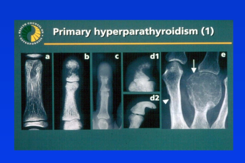
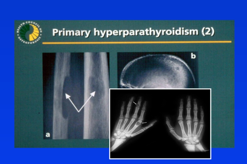
Слайд 19Bone and Joint Manifestations in Primary Hyperparathyroidism
Osteoporosis
Diffuse bone pain
Osteitis fibrosa cystica
Diffuse
Subperiostal bone resorption
Phalanges
Lamina dura
Distal clavicles
Cystic lesions
Brown tumors
Cysts
Deformities, Fractures, Pain
Arthritic symptoms
Resorption of articular bone
Periarticular metastatic calcification
Pseudogout
Gout


Слайд 24Other Considerations
Neuropsychological disturbances
Weakness and easy fatigability
Depression
Intellectual weariness
Increased sleep requirements
Improved by surgery
Onset
Increased bone loss


Слайд 2699mTc- sestamibi Parathyroid Scan
20 min
3 hours
90% sensitivity in parathyroid
tissue detection

Слайд 27What is 4D-CT?
4D-CT includes image sets in three planes (axial, coronal,
It is most commonly performed with three phases: non-contrast, arterial, and delayed phase imaging .
 from the")
Слайд 284D CT in diagnosis of PHPT
Arterial phase images are acquired 25


Слайд 30General Measures
Hydration
Adequate Mobility
Diet neither restrictive nor excessive in calcium
Adequate vitamin D
Prompt medical attention for the possibility of worsening of hypercalcemia (intercurrent illness accompanied by risk of dehydration)

Слайд 31Calcium-sensing Receptor
A member of the G protein-coupled receptor family
Contains seven
Large (~200 amino acids) cytosolic tail.

Слайд 32Inactivating Mutations in Calcium Sensing Receptor
Inactivating mutation
Familial hypocalciuric hypercalcemia (FHH) -
Calcium set point serum calcium
Urinary calcium reabsorption urinary
calcium
Neonatal severe hyperparathyroidism (NSHPT) – homozygous
parathyroid
Familial hypocalciuric hypercalcemia (FHH) - heterozygous
Calcium set point serum calcium
Urinary calcium reabsorption urinary
calcium
Neonatal severe hyperparathyroidism (NSHPT) – homozygous-incompatible with life if not resect the parathyroid
No need for parathyroid surgery in FHH!!
 - heterozygous Calcium set point")
Слайд 33Hypercalcemia of Malignancy
Lung, breast, and prostate cancer frequently invade skeleton and
Damage to skeleton usually late in course of disease
Bone damage associated with considerable worsening in patient’s quality of life
Multiple myeloma has skeletal complications in virtually 100% of cases
Breast and lung cancer also cause hypercalcemia of malignancy, without invading skeleton

Слайд 34PthrP Induced Hypercalcemia
Squamous cell carcinoma
Islet cell tumor (pancreas)
Adult T cell leukemia
Renal
Breast carcinoma
Adult T cell leukemiaRenal cell carcinomaBreast carcinoma")
Слайд 35PTHrP-PTH related peptide
Gen located on the chromosome 12
Gen is expressed in
Expression of the PTHrP gen in adult: plays role in breast development; presents in breast milk in high concentration
Mutation in embryo:
Heterozygous are normal
Homozygous –lethal mutation with major bone and cartilage abnormalities


Слайд 37Hydration
First step in the management of severe hypercalcemia - Isotonic saline
Usually
Hydration alone rarely leads to normalization in severe hypercalcemia
Rate of IV saline based on severity of hypercalcemia and tolerance of CVS for volume expansion, if possible achieve urine output of 300 ml/hr, that is then adjusted to maintain the urine output at 100 to 150 mL/hour.

Слайд 38Loop Diuretics
In the past intensive administration of furosemide was used (80
It needs aggressive fluid hydration (10 liters daily), saline therapy beyond that necessary to restore euvolemia.
Now use of calcitonin and bisphosphonates is more effective.

Слайд 39Calcitonin
The efficacy of calcitonin (4 IU/kg every 6-12 hours) is limited
Calcitonin and hydration provide a rapid reduction in serum calcium concentration, while a bisphosphonate provides a more sustained effect.
 is limited to the first 48")
Слайд 40Bisphosphonates
Structurally related to pyrophosphate
Great affinity for bone and their resistance to
Bind to hydroxyapatite in bone and inhibit the dissolution of crystals
Extremely long half life in bone
Poor GI absorption < 1%

Слайд 41Zoledronic Acid (ZOMERA)
Zoledronic acid belongs to a new class of highly
Heterocyclic, nitrogen-containing bisphosphonate composed of:
A core bisphosphonate moiety
An imidazole-ring side chain containing 2 critically positioned nitrogen atoms
Zoledronic acid belongs to a new class of highly potent bisphosphonatesHeterocyclic, nitrogen-containing bisphosphonate")
Слайд 42Denosumab Mechanism of Action
© 2007 Amgen. All rights reserved.
RANKL
RANK
OPG
Denosumab
Bone Formation
Hormones
Growth factors
Cytokines
Bone
Osteoclast Formation, Function,
and Survival Inhibited
CFU-GM
Pre-Fusion
Osteoclast
CFU-GM=colony forming unit granulocyte macrophage
Provided as an educational resource. Do not copy or distribute.
© 2007 Amgen. All rights reserved.
Osteoblasts
Calcium nadir 8-11 days after injection

Слайд 43Inhibit the growth of neoplastic lymphoid tissue
Counteract the effects of vitamin
Glucocorticoids


Слайд 45Hypocalcemia
Low PTH (hypoparathyroidism)
Genetic disorders: Abnormal parathyroid gland development
Abnormal PTH
Post-surgical
Autoimmune: Autoimmune polyglandular syndrome (associated with chronic mucocutaneous candidiasis and primary adrenal insufficiency)
Hypoparathyroidism due to activating antibodies to calcium-sensing receptor
Infiltration of the parathyroid gland (granulomatous, iron overload, metastases)
Radiation-induced destruction parathyroid glands
Hungry bone syndrome (post parathyroidectomy)
 Genetic disorders: Abnormal parathyroid gland development Abnormal PTH synthesis; Activating mutations of")
Слайд 46Hypocalcemia
High PTH (secondary hyperparathyroidism in response to hypocalcemia)
Vitamin D deficiency
Parathyroid hormone resistance: Pseudohypoparathyroidism
Hypomagnesemia
Renal disease
Loss of calcium from the circulation:
Hyperphosphatemia ; Tumor lysis ; Acute pancreatitis (the mechanism unknown)
Osteoblastic metastases; Acute respiratory alkalosis
Sepsis or severe burns- the cause appears to be a combination of impaired secretion of PTH coupled with reduced calcitriol production
 Vitamin D deficiency or resistance Parathyroid hormone")
Слайд 47Drugs induced hypocalcemia
Inhibitors of bone resorption (bisphosphonates, calcitonin, denosumab), especially in
Cinacalcet –calcimimetic
Calcium chelators (EDTA, citrate, phosphate) Foscarnet (due to intravascular complexing with calcium)
Phenytoin (due to conversion of vitamin D to inactive metabolites)
Fluoride poisoning
, especially in vitamin D deficiency Cinacalcet")
Слайд 48Trousseau's sign is the induction of carpopedal spasm by inflation of
Chvostek's sign is contraction of the ipsilateral facial muscles elicited by tapping the facial nerve just anterior to the ear
Hypocalcemia

Слайд 49Hypocalcemia: Carpopedal spasm and ECG changes
Carpopedal Spasm
Sinus rhythm with diffuse T
QT prolongation (The corrected qtc is 560 ms )
Prolongation is in the ST segment rather than the T waves

Слайд 50Treatment of Acute Hypocalcemia
Promptly correct symptomatic or severe hypocalcemia with cardiac
IV calcium therapy is suggested in asymptomatic patients with an acute decrease in serum corrected calcium to ≤7.5 mg/dL
(1 to 2 g of calcium gluconate, equivalent to 90 to 180 mg elemental calcium, in 50 mL of 5 percent dextrose) can be infused over 10 to 20 minutes. Is effective for 2-3 hours

Слайд 51Treatment of Acute Hypocalcemia
For those with milder symptoms of neuromuscular irritability
To effectively treat hypocalcemia in patients with concurrent magnesium deficiency,hypomagnesemia should be corrected first.
Identify and treat the cause of hypocalcemia and taper the infusion.
 and corrected serum")
Слайд 52To prevent Hypocalcemia due to Hungry bone syndrome
Start oral calcium and


Слайд 58Natpara
Natpara- for use in patients who
can’t be normalized on regular
oral therapy, or have complication
s due to it (nephrolithiasis, hyper
Calciuria)

Слайд 59Causes of magnesium depletion
Causes of magnesium depletion
Renal losses
Gasro-intestinal losses

Слайд 60Treatment of Hypomagnesemia
If the serum magnesium concentration is low, 2 g
Magnesium repletion should be continued as long as the serum magnesium concentration is less than 0.8 mEq/L (1 mg/dL or 0.4 mmol/L).
Persistent hypomagnesemia, as occurs in some patients with ongoing gastrointestinal (malabsorption) or renal losses, requires supplementation with oral magnesium, typically 300 to 400 mg daily divided into three doses.
 of magnesium")
Слайд 61Treatment of Hypomagnesemia
Oral repletion- if available and tolerable.
A typical daily
If a sustained-release preparation is not available, magnesium oxide 800 to 1600 mg (20 to 40 mmol [40 to 80 meq]) daily in divided doses may be used for moderate to severe hypomagnesemia.
Diarrhea frequently occurs with magnesium oxide therapy.