- Главная
- Разное
- Дизайн
- Бизнес и предпринимательство
- Аналитика
- Образование
- Развлечения
- Красота и здоровье
- Финансы
- Государство
- Путешествия
- Спорт
- Недвижимость
- Армия
- Графика
- Культурология
- Еда и кулинария
- Лингвистика
- Английский язык
- Астрономия
- Алгебра
- Биология
- География
- Детские презентации
- Информатика
- История
- Литература
- Маркетинг
- Математика
- Медицина
- Менеджмент
- Музыка
- МХК
- Немецкий язык
- ОБЖ
- Обществознание
- Окружающий мир
- Педагогика
- Русский язык
- Технология
- Физика
- Философия
- Химия
- Шаблоны, картинки для презентаций
- Экология
- Экономика
- Юриспруденция
Control of body temperature презентация
Содержание
- 1. Control of body temperature
- 2. Aims To understand the body’s control of
- 3. Thermoregulation The ability to keep the body
- 4. Temperature control Temperature control is the process
- 5. Temperature receptors in the skin detect changes
- 7. If we are too hot or too
- 9. Core temperature maintained despite of environmental changes
- 10. normal body temperature? Normal body temperature can
- 11. normal body rhythms
- 12. The average normal body temperature taken in
- 13. Causes of temperature variation Environmental Exercise Food/drink
- 14. An increase in body temperature can occur;
- 15. Considerations when taking a temperature Room temperature
- 16. Hyperthermia (heat stroke) Occurs when the body
- 17. fever Fever occurs when the core temperature
- 18. hypothermia Happens when the body temperature falls
- 19. Any questions?
- 20. Respiratory system Lynne Powell
- 21. Respiration is defined as; The transport of
- 22. Respiration v. ventilation Respiration is a chemical
- 24. The respiratory system The human respiratory system
- 26. Ventilation Movements of the ribs, rib muscles
- 27. Ventilation
- 28. Gas exchange We need to move oxygen
- 29. Gaseous exchange
- 30. Respirations are recorded for a number of
- 31. When measuring and recording breathing, the rate and pattern of breathing should be recorded.
- 32. The rate should be regular with equal
- 33. is the patient; mouth breathing
- 34. Average respiratory rates, by age: Average adult
- 35. Measuring respiratory rate The human respiration rate
- 36. PEAK FLOW MEASUREMENT Measurement of expiratory flow.
- 37. To take a peak flow reading you
- 38. Vitalograph
- 39. Infection control considerations DISPOSABLE MOUTH PIECES
- 40. ACTION PLAN FOR PATIENTS with asthma Green:
- 41. Pulse oximetry
- 42. Pulse oximetry Non-invasive method of monitoring the % of haemoglobin (Hb) saturated with oxygen.
- 43. Blood Red cells contain haemoglobin that have
- 44. Haemoglobin is the active oxygen carrying part
- 45. Most oxygen is carried by haemoglobin but
- 46. How does a pulse oximeter work? Calibrated
- 47. Limitations of pulse oximetry Not reliable in
- 48. Using an oximeter Resting readings should be
- 49. Results of pulse oximetry 95 - 100%
- 50. Medicines & healthcare products regulatory agency
- 51. Any questions?

Слайд 2Aims
To understand the body’s control of temperature and respiratory rate.
To look
at the anatomy and physiology relevant to temperature maintenance and respiration.
To discuss the normal values of temperature, respiratory rate and pulse oximetry.
To discuss factors that affect these normal values
To discuss the normal values of temperature, respiratory rate and pulse oximetry.
To discuss factors that affect these normal values

Слайд 3Thermoregulation
The ability to keep the body temperature within its limitations even
when the surrounding temperature is different.

Слайд 4Temperature control
Temperature control is the process of keeping the body at
a constant temperature of 37°C.
Our body can only stay at a constant temperature if the heat we generate is balanced and equal to the heat we lose.
Although our core temperature must be 37ºC, our fingers and toes can be colder. This is because energy is transferred from the blood as it travels to our fingers and toes.
Our body can only stay at a constant temperature if the heat we generate is balanced and equal to the heat we lose.
Although our core temperature must be 37ºC, our fingers and toes can be colder. This is because energy is transferred from the blood as it travels to our fingers and toes.

Слайд 5Temperature receptors in the skin detect changes in the external temperature.
They pass this information to the processing centre in the brain, called the hypothalamus.
The hypothalamus has temperature receptors to detect changes in the temperature of the blood.
the hypothalamus automatically triggers changes to effectors to ensure our body temperature remains constant, at 37°C.
The effectors are sweat glands and muscles.
The hypothalamus has temperature receptors to detect changes in the temperature of the blood.
the hypothalamus automatically triggers changes to effectors to ensure our body temperature remains constant, at 37°C.
The effectors are sweat glands and muscles.
Hypothalamus


Слайд 7If we are too hot or too cold, the processing centre
sends nerve impulses to the skin, which has two ways to either increase or decrease heat loss from the body's surface.
Tiny muscles in the skin can quickly pull the hairs upright to reduce heat loss, or lay them down flat to increase heat loss.
Hairs on the skin trap more warmth if they are standing up, and less if they are lying flat. Tiny muscles in the skin can quickly pull the hairs upright to reduce heat loss, or lay them down flat to increase heat loss.
If the body is too hot, glands in the skin secrete sweat onto the surface to increase heat loss by evaporation. This cools the body. Sweat secretion slows when the body temperature returns to normal.


Слайд 10normal body temperature?
Normal body temperature can vary slightly
It is influenced by
factors such as exercise, eating, sleeping and the time of the day & time of the month.
The lowest temperature is usually recorded at around 4am, the highest at 6 - 7pm.
The lowest temperature is usually recorded at around 4am, the highest at 6 - 7pm.


Слайд 12The average normal body temperature taken in the mouth is 37ºC
(98.6ºF), but anywhere between 36.5ºC and 37.2ºC (97.7ºF and 99ºF) may be normal.
The accepted range of "normal" temperature is from 97F (36.1C) to 99F (37.2C)
For
Rectal = 34.4–37.8 °C (94–100 °F)
Tympanic cavity = 35.4–37.8 °C (96–100 °F)
Auxiliary = 35.5–37.0 °C (96–99 °F)
The accepted range of "normal" temperature is from 97F (36.1C) to 99F (37.2C)
For
Rectal = 34.4–37.8 °C (94–100 °F)
Tympanic cavity = 35.4–37.8 °C (96–100 °F)
Auxiliary = 35.5–37.0 °C (96–99 °F)
Normothermia/euthermia
, but anywhere between")
Слайд 13Causes of temperature variation
Environmental
Exercise
Food/drink
Dehydration (vomiting & diarrhoea)
Drugs
Infection
Inflammation
Disease
DrugsInfection InflammationDisease")
Слайд 14An increase in body temperature can occur;
Infective conditions
Inflammation
Immunological diseases
Lupus
Sarcoidosis
Inflammatory bowel disease
Drugs
Adverse
reaction to drugs/immunisation
Chemotherapy
Recreational drug withdrawal
Metabolic disorders
Gout (local)
hyperthyroidism
Chemotherapy
Recreational drug withdrawal
Metabolic disorders
Gout (local)
hyperthyroidism

Слайд 15Considerations when taking a temperature
Room temperature
How is the patient dressed
Temperature site
The
equipment
Is the equipment in working order
Why are you taking the temperature
Any medical history
Is the equipment in working order
Why are you taking the temperature
Any medical history

Слайд 16Hyperthermia (heat stroke)
Occurs when the body produces or absorbs more heat
the it can dissipate.
The most common cause is heat stroke and adverse reactions to drugs (cancer treatment eg radiotherapy)
The body’s set temperature remains the same
The most common cause is heat stroke and adverse reactions to drugs (cancer treatment eg radiotherapy)
The body’s set temperature remains the same
Occurs when the body produces or absorbs more heat the it can dissipate.The")
Слайд 17fever
Fever occurs when the core temperature is set higher
Usually in response
to bacterial or viral infections
Certain cells in the blood release pyrogens (a substance that induces fever) which have a direct effect on the hypothalamus causing body temperature to raise
Certain cells in the blood release pyrogens (a substance that induces fever) which have a direct effect on the hypothalamus causing body temperature to raise

Слайд 18hypothermia
Happens when the body temperature falls below 35c
Hypothermia can quickly become
life threatening and treated as a medical emergency
Usually triggered by being in a cold environment
The elderly, the ill and those who are unable to move around easily to generate heat are most at risk
Babies are also prone to hypothermia because their ability to control temperature isn’t fully developed
Usually triggered by being in a cold environment
The elderly, the ill and those who are unable to move around easily to generate heat are most at risk
Babies are also prone to hypothermia because their ability to control temperature isn’t fully developed


Слайд 21Respiration is defined as;
The transport of oxygen from the outside air
to the cells within tissues and the transport of carbon dioxide in the other direction

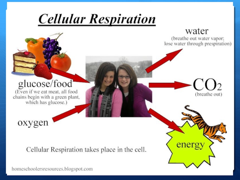
Слайд 22Respiration v. ventilation
Respiration is a chemical reaction that happens in all
living cells. It is the way that energy is released from glucose, for our cells to use to keep us functioning.
Remember that respiration is not the same as breathing (which is properly called ventilation).
Breathing includes inspiration (breathing in) and exhalation (breathing out)
Remember that respiration is not the same as breathing (which is properly called ventilation).
Breathing includes inspiration (breathing in) and exhalation (breathing out)

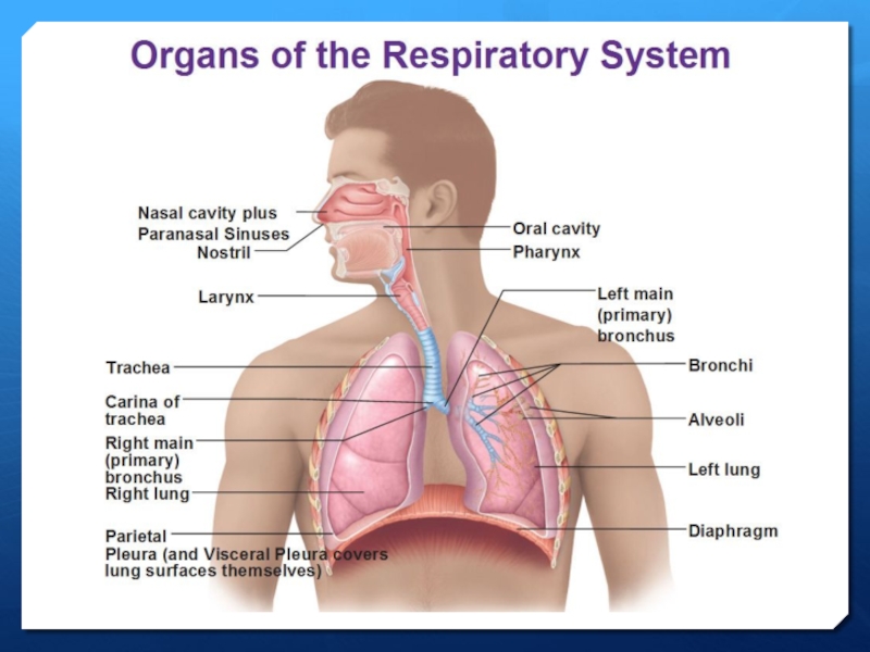
Слайд 24The respiratory system
The human respiratory system contains the organs that allow
us to get the oxygen we need and to remove the waste carbon dioxide we don't need. It contains these parts:
lungs
tubes leading from the lungs to the mouth and nose
various structures in the chest that allow air to move in and out of the lungs. The diaphragm, ribs,
lungs
tubes leading from the lungs to the mouth and nose
various structures in the chest that allow air to move in and out of the lungs. The diaphragm, ribs,

Слайд 26Ventilation
Movements of the ribs, rib muscles and diaphragm allow air into
and out of the lungs.
this is called breathing or ventilation, not respiration.
Air passes between the lungs and the outside of the body through the trachea
The trachea divides into two bronchi, with one bronchus for each lung.
Each bronchus divides further in the lungs into smaller tubes called bronchioles.
At the end of each bronchiole, there is a group of tiny air sacs. These air sacs have bulges called alveoli


Слайд 28Gas exchange
We need to move oxygen from the air into the
blood and waste carbon dioxide from the blood into the air
alveoli are adapted to make gas exchange in lungs happen easily and efficiently
alveoli are adapted to make gas exchange in lungs happen easily and efficiently


Слайд 30Respirations are recorded for a number of reasons:
To acquire a baseline.
To
monitor a patient with breathing problems.
To aid in the diagnosis of disease.
To evaluate the response to medication that affects the respiratory system.
To aid in the diagnosis of disease.
To evaluate the response to medication that affects the respiratory system.

Слайд 31When measuring and recording breathing, the rate and pattern of breathing
should be recorded.

Слайд 32The rate should be regular with equal pause between each breath.
The rate can be irregular with disease of the respiratory system. Any irregularities should be noted and reported.
When observing the respiratory rate, noting the colour of the patients lips is important. They may be cyanosed (blue) or discoloured if the patient has respiratory problems. Cyanosis can also be observed in the nail bed, tip of the nose and ear lobes.
The patient’s oxygen saturation (SaO2) may be recorded using a pulse oximeter.
Pulse oximetry provides a reading oxygenation in the red blood cells. Using a pulse oximeter may require the patient to have less arterial blood gases performed, by providing the medical team with a guide to the patients oxygenation level.

Слайд 33is the patient;
mouth breathing
pursing the lips on expiration,
using the
abdominal muscles
flaring the nostrils
Observe
Lips
Ear lobes
Tip of the nose
flaring the nostrils
Observe
Lips
Ear lobes
Tip of the nose
Observing breathing

Слайд 34Average respiratory rates, by age:
Average adult 10-20 breaths/min
Newborns: Average 44 breaths
per minute
Infants: 20–40 breaths per minute
Preschool children: 20–30 breaths per minute
Older children: 16–25 breaths per minute
Adults during strenuous exercise 35–45 breaths per minute
Athletes' peak 60–70 breaths per minute
Infants: 20–40 breaths per minute
Preschool children: 20–30 breaths per minute
Older children: 16–25 breaths per minute
Adults during strenuous exercise 35–45 breaths per minute
Athletes' peak 60–70 breaths per minute

Слайд 35Measuring respiratory rate
The human respiration rate is usually measured when a
person is at rest.
Record the number of breaths for one full minute by counting how many times the chest rises.
When checking respiration, it is important to also note whether a person has any difficulty breathing.
The invasiveness of touch/observation is enough to sometimes make significant changes in breathing. .
Record the number of breaths for one full minute by counting how many times the chest rises.
When checking respiration, it is important to also note whether a person has any difficulty breathing.
The invasiveness of touch/observation is enough to sometimes make significant changes in breathing. .

Слайд 36PEAK FLOW MEASUREMENT
Measurement of expiratory flow.
Standard range peak flow meters are
suitable for both adults and children.
Low range peak flow meters are designed for adults and children with severely impaired function of the lungs.
Readings will vary according to age, height and gender (male or female).
Record children’s height at every visit (at least annually).
Low range peak flow meters are designed for adults and children with severely impaired function of the lungs.
Readings will vary according to age, height and gender (male or female).
Record children’s height at every visit (at least annually).

Слайд 37To take a peak flow reading you should:
check that the pointer
is at zero.
preferably stand or sit in a comfortable, upright position.
hold the peak flow meter level (horizontally) and keep your fingers away from the pointer.
take a deep breath and close your lips firmly around the mouthpiece.
then blow as hard as you can - as if you were blowing out candles on a birthday cake - remember it is the speed of your blow that is being measured.
look at the pointer and check your reading.
reset the pointer back to zero.
do this three times and record the highest reading in your daily asthma diary.


Слайд 40ACTION PLAN FOR PATIENTS with asthma
Green: 80 to 100 percent of
your personal best peak flow measurement; asthma is under control.
Yellow: 50 to 79 percent of your personal best peak flow measurement; asthma is getting worse; you may need to use quick-relief medications or other medication, as directed by your doctor.
Red: below 50 percent of your personal best peak flow measurement; medical alert, take quick-relief medication and seek medical help immediately.


Слайд 42Pulse oximetry
Non-invasive method of monitoring the % of haemoglobin (Hb) saturated
with oxygen.
 saturated with oxygen.")
Слайд 43Blood
Red cells contain haemoglobin that have the ability to pick up
and release oxygen under differing pressures

Слайд 44Haemoglobin is the active oxygen carrying part of the erythrocyte (red
blood cell)
Blood carries oxygen in 2 ways – dissolved in plasma (approx 3%) & attached to haemoglobin
Blood carries oxygen in 2 ways – dissolved in plasma (approx 3%) & attached to haemoglobin
Blood carries oxygen")
Слайд 45Most oxygen is carried by haemoglobin but 3 factors influence the
amount of oxygen delivered to the tissue;
Tissue perfusion – blood supply to area
Amount of haemoglobin
Saturation of haemoglobin
Tissue perfusion – blood supply to area
Amount of haemoglobin
Saturation of haemoglobin

Слайд 46How does a pulse oximeter work?
Calibrated during manufacture.
It emits two wavelengths
which are partly absorbed by haemoglobin.
It is able to distinguish between the types of blood vessels and pick up arterial flow (pulsitile).
It absorbs the red and infrared light of the systolic component of the wavelength.
It then computes the amount of haemoglobin that is saturated with oxygen.
It is able to distinguish between the types of blood vessels and pick up arterial flow (pulsitile).
It absorbs the red and infrared light of the systolic component of the wavelength.
It then computes the amount of haemoglobin that is saturated with oxygen.

Слайд 47Limitations of pulse oximetry
Not reliable in patient’s with poor circulation (e.g.
peripheral vascular disease, anaemia, hypothermia, critically ill pts)
Does not work through nail vanish, dyes or pigments.
Not reliable in patients that have an irregular pulse rate.
Shivering and movement give false readings.
There have been reports of skin burns (earlier models).
Not reliable in bright overhead lighting.
Extremely misleading in cases of carbon monoxide poisoning (causes saturation levels toward 100%) CO-oximetry should be used.

Слайд 48Using an oximeter
Resting readings should be taken for at least 5
mins
The sensor is usually placed on a thin part of the patient’s body e.g. fingertip, earlobe
The hand should be rested either on the chest at the level of the heart or on a flat surface.
Ensure the digit is inserted fully as light should not reach the detector except through the tissue.
Check that the displayed heart rate correlates to a manually checked rate

Слайд 49Results of pulse oximetry
95 - 100% = Acceptable normal ranges
92%
or less = Refer for oxygen assessment
88 - 94% = Hypoxic drive problem
100% = may indicate carbon-monoxide poisoning or cyanide poisoning
88 - 94% = Hypoxic drive problem
100% = may indicate carbon-monoxide poisoning or cyanide poisoning