- Главная
- Разное
- Дизайн
- Бизнес и предпринимательство
- Аналитика
- Образование
- Развлечения
- Красота и здоровье
- Финансы
- Государство
- Путешествия
- Спорт
- Недвижимость
- Армия
- Графика
- Культурология
- Еда и кулинария
- Лингвистика
- Английский язык
- Астрономия
- Алгебра
- Биология
- География
- Детские презентации
- Информатика
- История
- Литература
- Маркетинг
- Математика
- Медицина
- Менеджмент
- Музыка
- МХК
- Немецкий язык
- ОБЖ
- Обществознание
- Окружающий мир
- Педагогика
- Русский язык
- Технология
- Физика
- Философия
- Химия
- Шаблоны, картинки для презентаций
- Экология
- Экономика
- Юриспруденция
Clinical anatomy of abdominal cavity презентация
Содержание
- 1. Clinical anatomy of abdominal cavity
- 2. liver in the upper right quadrant of
- 3. borders: superior: inferior surface of diaphragm Inferior:
- 4. Borders: Superior: mesocolon transversum Inferior: inlet of
- 5. After cutting through the abdominal wall, if
- 6. lig. falciforme lig. coronarium hepatis lig. triangulare
- 7. duodenojejunal recess superior ileocaecal recess inferior ileocaecal
- 8. Plica gastropancreatica Plica ileocecalis Plica duodenalis superior
- 9. RIGHT MESENTERIC SINUS borders: medial-root of
- 10. Right paracolic canal communicates with right hepatic
- 11. HEPATIC BURSA Borders: Superior – diaphragm Inferior
- 12. Pregastric bursa Borders: Anterior – left lobe
- 13. BORDERS: Superior – lobus caudatus hepatis Inferior
- 14. The branches to the stomach arise from
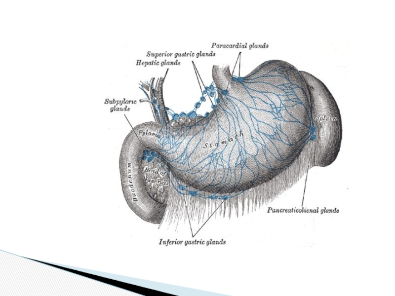
- 15. The stomach drains either directly or indirectly
- 16. Nerve supply
- 18. Gastritis (acute or stress) Produces inflammation
- 19. Menetrier’s Disease (aka Hypertrophic Gastritis)
- 20. Gastric Polyps
- 21. Bezoars
- 22. The “Culprit” H. pylori Treatment: Triple therapy
- 23. Gastric ulcers
- 24. Gastric Ulcers
- 25. History of Peptic Ulcer Surgery Harberer 1882-
- 26. Laser Coagulation of Bleeding Ulcer
- 27. Coil Embolization of Bleeding Ulcer
- 28. Pyloroplasty for Bleeding Ulcer
- 29. Open Surgical Procedures Truncal vagotomy and pyloroplasty
- 30. GASTROSTOMY Temporary gastrostomy Minimal gastrostomy Vitzel’s gastrostomy
- 32. Roux -en -Y Reconstruction
- 33. Antecolic and Retrocolic BII
- 34. Truncal Vagotomy Resect 1-2cm of each vagal
- 35. Antrectomy and Truncal Vagotomy with BI
- 36. Truncal Vagotomy and Antrectomy Entails distal gastrectomy
- 37. Selective Vagotomy Total denervation of the stomach
- 38. Highly Selective Vagotomy Spares nerves of Latarjet,
- 39. Types of Vagotomies
- 40. Gastric Adenocarcinoma
- 41. Duodenum 4 parts Metabolically active Produces
- 42. Duodenum Brunner’s glands Blood supply: GDA- superior pancreaticoduodenal SMA- inferior pancreaticoduodenal
- 43. Blood Supply of the Duodenum superior pancreaticoduodenal
- 44. Duodenal Ulcers
- 45. Obstruction
- 46. Small Bowel Obstruction History Prior surgery Hernias
- 47. Intestinum Crasum
- 48. Large Bowel Obstruction
- 49. colostomy
- 50. Anastamosis Stapled vs. Hand-Sewn Brundage et al.
- 51. Anastamosis Burch et al. Ann of Surg.
- 52. Appendix vermiformis
- 54. The caecum was at McBurney's point in
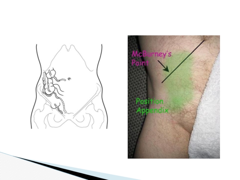
- 55. Topography of appendix vermiformis and ceacum
- 57. Ulcerative Colitis Disease Severity Mild colitis: 20%
- 58. Subtotal Colectomy
- 59. Liver
- 60. Liver
- 61. Mosby items and derived items © 2006 by Mosby, Inc. Slide Liver Structure
- 62. Porto-caval anastomoses
- 63. Caput Medusa
- 65. Varices on EGD
- 66. Varix Banding
- 67. Gall bladder
- 68. Arteries of the gall bladder
- 69. Innervation of gall bladder
- 70. Lymphatic drainage of the gallbladder
- 71. Harvest Time
- 72. CT Scan
- 73. Plain Films
- 74. Ultrasound
- 75. Laparoscopic Cholecystectomy
- 76. cancer
- 77. Surgical Options Simple cholecystectomy Radical cholecystectomy Radical

Слайд 2liver in the upper right quadrant of the cavity. It is
the tip of the gall bladder (gb) hanging down under the margin of the liver
stomach (st) in the upper left quadrant
a small edge of the spleen (sp) in the upper left quadrant
greater omentum (go) covering most of the abdominal structures
small intestines (ileum) (il) in the lower right quadrant
sometimes the transverse colon (tc) can be seen through a thin portion of the greater omentum.
Abdomilal cavity

Слайд 3borders:
superior: inferior surface of diaphragm
Inferior: mesocolon transversum
Contents: hepatic bursa, pregastric bursa,
Upper storey

Слайд 4Borders:
Superior: mesocolon transversum
Inferior: inlet of the lesser pelvis
contents:
Right & left paracolic
Right & left mesenteric sinuses
Mesentry
Sigmoid mesocolon
Duodenojejunal recess
Superior and inferior ileocaecal recesses
Large and small intestines
Inferior storey

Слайд 5After cutting through the abdominal wall, if you put your hand
From here you follow the mesentery of the small intestine (7) going around its coils until you reach the other side of the mesentery back down to the posterior abdominal wall where you will cross over the horizontal part of the duodenum (8). Your finger will then travel along the inferior aspect of the gastrocolic ligament (9), down the posterior surface of the greater omentum (go) to its lower border and back up along its anterior surface(11). Your finger then passes over the anterior surface of the stomach (12), along the anterior lamina of the lesser omentum (13). At this time you probably couldn't continue the trip because you would have to enter the epiploic foramen (ef) to enter the lesser peritoneal cavity (lpc) where visceral peritoneum lines this space anteriorly and parietal peritoneum posteriorly.
peritoneum

Слайд 6lig. falciforme
lig. coronarium hepatis
lig. triangulare
lig. hepatogastricum
lig. hepatoduodenale
lig. hepatocolicum
lig. hepatorenale
lig. gastrophrenicum
lig. gastrolienale
lig.
lig. gastropancreaticum
lig. phrenicoesophageale
lig. phrenicocolicum
lig. phrenicorenale
lig. phrenicolienale
lig. pancreaticolienale
lig. lienorenale
lig. pyloropancreaticum
lig. duodenorenale
ligaments

Слайд 7duodenojejunal recess
superior ileocaecal recess
inferior ileocaecal recess
retrocaecal recess
intersigmoid recess
Recesses - pouches formed

Слайд 8Plica gastropancreatica
Plica ileocecalis
Plica duodenalis superior
Plica duodenalis inferior
Plica umbilicalis mediana
Plica umbilicalis medialis
Plica
Folds – reflection of the peritoneum arised from the abdominal wall by uderlying structures

Слайд 9RIGHT MESENTERIC SINUS
borders:
medial-root of the mesentry
Lateral – ascending colon
Superior –
LEFT MESENTERIC SINUS
Borders
Medial – descending colon
Lateral – root of the mesentry
Inferior – sigmoid colon
sinuses

Слайд 10Right paracolic canal communicates with right hepatic bursa
Borders:
Medial – ascending colon
Lateral
inferior – caecum
Left paracolic canal communicates with lesser pelvis
Borders:
Medial – descending colon
Lateral – parietal peritoneum
Superior – phrenicocolic ligament
Paracolic canals

Слайд 11HEPATIC BURSA
Borders:
Superior – diaphragm
Inferior – transverse mesocolon
Anterior – anterior abdominal wall
Medial
Pathology: abscess from the inferior storey of the abdominal cavity may spread here and cause subphrenic abscess through the right paracolic canal
Bursae of the abdominal cavity

Слайд 12Pregastric bursa
Borders:
Anterior – left lobe of the liver and anterior abdominal
Posterior – lesser omentum
Pathology: abscess from this bursa may spread to the omental bursa
Bursae of the abdominal cavity

Слайд 13BORDERS:
Superior – lobus caudatus hepatis
Inferior – mesocolon transversum
Anterior – stomach &
Posterior – parietal peritoneum
Pathology: inflammation from this bursa may spread to the general peritoneal cavity through the epiploicc foramen.
FORAMEN EPIPLOICUM
BORDERS
Superior – lobus caudatus hepatis
Inferior – superior part of duodenum
Anterior – lig.hepatoduodenale
Posterior – lig.hepatorenale, parietal peritoneum which covers v.cava inferior
Omental bursa (bursa omentalis)

Слайд 14The branches to the stomach arise from the above: celiac (C)
left gastric (LG) -
esophageal (E)
splenic (S) which gives rise to:
short gastric (SG) - supplies area of the fundus
left gastroepiploic (LGE) - supplies the left part of greater curvature of the stomach
common hepatic (CH)
gastroduodenal (GD)
right gastric (RG) - supplies right side of lesser curvature of the stomach
right gastroepiploic (RGE) - supplies the right part of the greater curvature of the stomach
stomach
left gastric (LG) - supplies the lesser curvature")
Слайд 15The stomach drains either directly or indirectly into the portal vein
left gastroepiploic (LGE) along greater curvature to superior mesenteric vein (SM)
right gastroepiploic (RGE) from the right end of greater curvature to superior mesenteric vein (SM)
left gastric vein (LG) from the lesser curvature of the stomach to the portal vein (PV)
right gastric vein (RG) from the lesser curvature of the stomach to the portal vein (PV)
Venous drainage from stomach
")

Слайд 18Gastritis (acute or stress)
Produces inflammation of the mucosa.
Can be associated with
Causes:
H. pylori, NSAIDS, bile reflux, Etoh, radiation, local trauma, physiologic stress.
Produces inflammation of the mucosa.Can be associated with erosions and bleeding.Causes:H. pylori,")
")


Слайд 25History of Peptic Ulcer Surgery
Harberer 1882- first gastric resection for ulcer
Billroth
Hofmeister 1896- Retrocolic anastamosis
Dragstedt 1943- Truncal vagotomy
Visick 1948- vagotomy and drainage
Johnson 1970- highly selective vagotomy


Слайд 29Open Surgical Procedures
Truncal vagotomy and pyloroplasty
Truncal vagotomy and gastrojejunostomy
Truncal vagotomy and
Highly selective vagotomy

Слайд 30GASTROSTOMY
Temporary gastrostomy
Minimal gastrostomy
Vitzel’s gastrostomy
Stamm-Kader’s gastrostomy
Permanent gastrostomy
Toprover’s gastrostomy
Beck Jian’s gastrostomy
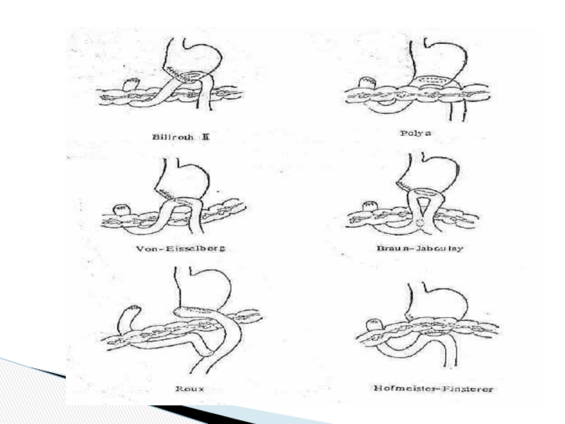
PARTIAL RESECTION OF
Billroth I – the stump of the stomach is anastomosed with that of the duodenum
Billroth II - the stump of the stomach is anastomosed with the initial portion of the ileum
Modifications of Billroth II
Operations on stomach


Слайд 34Truncal Vagotomy
Resect 1-2cm of each vagal trunk on distal esophagus.
Reduces acid
Denervates parietal cells, antral pump, pyloric sphincter mechanism.
Delays gastric emptying, so need drainage.
With pyloroplasty recurrence 3-10%
With pyloroplasty morbidity 1-2%


Слайд 36Truncal Vagotomy and Antrectomy
Entails distal gastrectomy of 50-60% of stomach.
Removes parietal
Requires a BI or BII reconstruction.
Recurrence rate 0.6-4%
Morbidity rate 0.9-1.6%

Слайд 37Selective Vagotomy
Total denervation of the stomach from diaphragmatic crus to pylorus.
Procedure

Слайд 38Highly Selective Vagotomy
Spares nerves of Latarjet, but divides vagal branches to
Antral innervation is thus preserved, gastric emptying preserved, so drainage procedure unnecessary.
Recurrence rate 10-15%
Lowest morbidity of all


Слайд 41Duodenum
4 parts
Metabolically active
Produces many enzymes
D2: site of pacemaker
D2: posterolateral insertion of
Becomes jejunum at the _____________?

Слайд 42Duodenum
Brunner’s glands
Blood supply:
GDA- superior pancreaticoduodenal
SMA- inferior pancreaticoduodenal

Слайд 43Blood Supply of the Duodenum
superior pancreaticoduodenal
anterior and posterior branches
inferior pancreaticoduodenal
anterior and
duodenum


Слайд 46Small Bowel Obstruction
History
Prior surgery
Hernias
Signs and Symptoms
Colicky abdominal pain
Nausea and vomiting
Abdominal distension
Rectal
No peritoneal signs


Слайд 50Anastamosis
Stapled vs. Hand-Sewn
Brundage et al. J trauma. 1999
Multicenter retrospective cohort design
“anastamotic

Слайд 51Anastamosis
Burch et al. Ann of Surg. 1999.
Prospective randomized trial of single-layer
NB: Important to invert, 4-6mm seromuscular bites, 5mm advances, larger bites at mesenteric border
Single layer – similar leak rate (approx 2%), cheaper, faster
Burch et al. Ann Surg. 1999


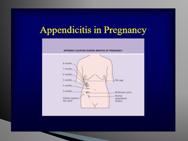
Слайд 54The caecum was at McBurney's point in 245 (80.9%) patients, pelvic
The average length was 8.9 cm in males and 9.4 cms in females. The appendix was commonly found to be retrocaecal (58.3%) on pelvic (21.7%) or paracaecal (11.7%). Anomalies of the appendix were more common in children than adults and occurred in 47% of cases.
 patients, pelvic in 45 (14.9%) and")

Слайд 57Ulcerative Colitis
Disease Severity
Mild colitis: 20%
Moderate colitis: 71%
Severe colitis: 9%
Acute disease complications
Toxic
Perforation
Hemorrhage
Langholz 1991


Слайд 77Surgical Options
Simple cholecystectomy
Radical cholecystectomy
Radical chole w/ anatomic liver resection
Radical chole w/