- Главная
- Разное
- Дизайн
- Бизнес и предпринимательство
- Аналитика
- Образование
- Развлечения
- Красота и здоровье
- Финансы
- Государство
- Путешествия
- Спорт
- Недвижимость
- Армия
- Графика
- Культурология
- Еда и кулинария
- Лингвистика
- Английский язык
- Астрономия
- Алгебра
- Биология
- География
- Детские презентации
- Информатика
- История
- Литература
- Маркетинг
- Математика
- Медицина
- Менеджмент
- Музыка
- МХК
- Немецкий язык
- ОБЖ
- Обществознание
- Окружающий мир
- Педагогика
- Русский язык
- Технология
- Физика
- Философия
- Химия
- Шаблоны, картинки для презентаций
- Экология
- Экономика
- Юриспруденция
Cardiovascular system. Systolic blood pressure презентация
Содержание
- 1. Cardiovascular system. Systolic blood pressure
- 2. Plan of the lectures Semiotic of disorders of CVS.
- 3. BLOOD PRESSURE Systolic blood pressure:
- 4. COMPLAINS Weakness Fatigability Disorders of appetite Dyspnoea
- 5. INSPECTION General condition Position of child
- 6. PALPATION Localization of apical thrust Cardiac
- 7. Estimation of pulse: Rhythm (N - respiratory
- 8. PERCUSSION Method of percussion of children over
- 9. BORDERS OF RELATIVE HEART’S DULNESS
- 10. BORDERS OF RELATIVE HEART’S DULNESS
- 11. Right under 2
- 12. apical thrust 4 intercostals space to the
- 13. Left under 2
- 14. SEQUENCE OF AUSCULTATION OF HEART
- 15. 1st Cardiac apex
- 16. 2nd II intercostals space to the right of sternum
- 17. 3rd II intercostals space to the left of sternum
- 18. 4th Place of connection xiphisternum to sternum, a little to the right
- 19. 5th Place of joining III-IV ribs to the edge of sternum
- 20. CARDIAC MURMURS
- 21. Cardiac murmurs – additional sounds heard during auscultation of the heart.
- 22. CHARACTERISTIC OF MURMURS Organic Functional Organic-functional Physiological
- 24. RHEUMATIC FEVER (ACUTE RHEUMATIC HEART DISEASE)
- 25. Diagnosis Diagnosis of ARF is
- 26. SYMPTOMS Carditis: chest pain, shortness or breath,
- 27. SIGNS carditis tachycardia murmur: mitral insufficiency, relative
- 28. INVESTIGATIONS Chest radiography: cardiomegaly when carditis is
- 29. Modified Jones criteria for the diagnosis of acute rheumatic fever
- 30. MYOCARDITIS Definition Inflammation of myocardium in association
- 31. SYMPTOMS Poor feeding or decreased appetite, shortness
- 32. SIGNS Tachycardia, irregular pulse. Soft, indistinct heart
- 33. INVESTIGATIONS ECG Chest radiography Echocardiography (ECHO) Radionuclide studies Laboratory studies Endomyocardial biopsy
- 34. ENDOCARDITIS
- 35. SYMPTOMS Fever and sweating. Easy fatiguability, malaise.
- 36. INVESTIGATIONS Laboratory tests Blood cultures. Acute phase reactants Complete blood count Urinanalysis: hematuria. Echocardiography!
- 37. COMPLICATIONS Destruction of cardiac valve tissue: can
- 38. PERICARDITIS AND TAMPONADE
- 39. Pericarditis SYMPTOMS AND SIGNS Mild to severe
- 40. Tamponade Symptoms similar to pericarditis: pain, cough,
- 41. INVESTIGATIONS • For
- 42. Pericarditis Complete blood count, erythrocyte sedimentation
- 43. Tamponade Echocardiography. Complications -Relapsing or constrictive
- 44. CONGENITAL HEART DISEASE
- 45. A ventricular septal defect Oxygenated blood is
- 47. AN ATRIAL SEPTAL DEFECT Oxygenated blood
- 49. A PATENT DUCTUS ARTERIOSUS Oxygenated blood from
- 51. TETRALOGY OF FALLOT (1) Right ventricular outflow
- 53. COARCTATION OF THE AORTA Narrowing of


Слайд 3BLOOD PRESSURE
Systolic blood pressure:
till 1 year old =
76 + 2 x n (n – months)
above 1 year old = 90 + 2 x n (n – years old)
Diastolic blood pressure:
above 1 year old = 60 + n (n – years old)
above 1 year old = 90 + 2 x n (n – years old)
Diastolic blood pressure:
above 1 year old = 60 + n (n – years old)

Слайд 4COMPLAINS
Weakness
Fatigability
Disorders of appetite
Dyspnoea
Color of the skin: cyanosis or paleness
Pain of the
heart region
Headache
Palpitation
Headache
Palpitation

Слайд 5INSPECTION
General condition
Position of child in the bed
Physical development (proportional or disproportional)
Deformation
on the chest (cardiac hump)
Color of the skin
Temperature of extremities
Moistness of the skin
Edema of low extremities
Pulsation of carotids
Jugular venous distention
Visible pulsation of heart
(intensive apical thrust (N-1cm2); heart thrust)
Color of the skin
Temperature of extremities
Moistness of the skin
Edema of low extremities
Pulsation of carotids
Jugular venous distention
Visible pulsation of heart
(intensive apical thrust (N-1cm2); heart thrust)
Deformation on the chest")
Слайд 6PALPATION
Localization of apical thrust
Cardiac thrust
Trembling above the mitral valve and it’s
characteristic
Presents of cat’s purr
Presents of cat’s purr

Слайд 7Estimation of pulse:
Rhythm (N - respiratory arrhythmia reveals from 2 till
10 years old)
Rate
Symmetric
Synchronic
Tension
Size
Rate
Symmetric
Synchronic
Tension
Size
 Rate Symmetric Synchronic Tension Size")
Слайд 8PERCUSSION
Method of percussion of children over 4 years old is same
with adults.
Until 4 years old use modification.
Until 4 years old use modification.


Слайд 10BORDERS OF RELATIVE HEART’S DULNESS
Upper
under
2 у.о. II costae
2 – 6 II intercostals space
7 – 12 upper border of III costae
older 12 III costae
or
III intercostals space
2 – 6 II intercostals space
7 – 12 upper border of III costae
older 12 III costae
or
III intercostals space

Слайд 11Right
under 2
2 cm outside
from
right sternalis line
2 – 6
1 cm outside from
right sternalis line
7 – 12
0,5 cm outside from
right sternalis line
older 12
right sternalis line
right sternalis line
2 – 6
1 cm outside from
right sternalis line
7 – 12
0,5 cm outside from
right sternalis line
older 12
right sternalis line

 from left medioclavicular line")
Слайд 13Left
under 2
2 cm outside from
left medioclavicular line
2 – 6 1 cm outside from
left medioclavicular line
7 – 12 0,5 cm outside from
left medioclavicular line
older 12 left medioclavicular line or 0,5 cm inwards
left medioclavicular line
2 – 6 1 cm outside from
left medioclavicular line
7 – 12 0,5 cm outside from
left medioclavicular line
older 12 left medioclavicular line or 0,5 cm inwards


Слайд 22CHARACTERISTIC OF MURMURS
Organic
Functional
Organic-functional
Physiological
Cardiac
Extracardial
Combined
Systolic
Diastolic
Timbre
Irradiation
Force
Duration


Слайд 24RHEUMATIC FEVER
(ACUTE RHEUMATIC HEART DISEASE)
Rheumatic heart disease (RHD) occurs after
acute rheumatic fever (ARF), which is a postinfectious immune disease secondary to a streptococcal infection.
RHD affects the valves of the heart, with the mitral and aortic being the most commonly affected valves. RHD is more likely to occur with recurrent episodes.
RHD affects the valves of the heart, with the mitral and aortic being the most commonly affected valves. RHD is more likely to occur with recurrent episodes.
Rheumatic heart disease (RHD) occurs after acute rheumatic fever (ARF),")
Слайд 25Diagnosis
Diagnosis of ARF is made from the modified Jones criteria,
with a positive diagnosis requiring two major or one major and two minor criteria.

Слайд 26SYMPTOMS
Carditis: chest pain, shortness or breath, cough, palpitations with sensation of
elevated or irregular rate or rhythm, anorexia, fatigue, and exercise intolerance.
Arthritis: occurs in 70% of patients with ARF; symptoms include joint pain.
Sydenham’s chorea: affects 15% of patients with ARF; reflects involvement of basal ganglia of central nervous system; appears ~3 months after streptococcal infection; symptoms include emotional liability and loss of attention span.

Слайд 27SIGNS
carditis
tachycardia
murmur: mitral insufficiency, relative mitral stenosis, or aortic insufficiency
congestive heart failure
arrhythmias
pericarditis
arthritis: large joints (knees, ankles, wrists, elbows); asymmetric and migratory
Sydenham’s chorea: involuntary and purposeless movements and muscular incoordination; erythema marginatum: 5%; subcutaneous nodules: <5%.

Слайд 28INVESTIGATIONS
Chest radiography: cardiomegaly when carditis is present; pulmonary venous congestion and/or
pulmonary edema may be present when congestive heart failure occurs.
ECG: sinus tachycardia; PR prolongation with myocarditis; nonspecific ST-T abnormalities or T wave inversion; premature atrial or ventricular contractions.
Echocardiography: used to evaluate the presence and degree of myocardial dysfunction, specific chamber enlargement
Acute phase reactants: elevated WBC , ESR, CRP.
Streptococcal infection: elevated streptozyme; anti-streptolysin O, antistreptokinase, anti-hyaluronidase.


Слайд 30MYOCARDITIS
Definition
Inflammation of myocardium in association with necrosis.
Presentation
Mild to moderate congestive heart
failure (CHF).
Severe CHF or shock.
Non-CHF: arrhythmia, sudden death.
History of prior gastrointestinal or flu-like illness.
Severe CHF or shock.
Non-CHF: arrhythmia, sudden death.
History of prior gastrointestinal or flu-like illness.
.Severe CHF or")
Слайд 31SYMPTOMS
Poor feeding or decreased appetite, shortness of breath or dyspnea, easy
fatigability or general malaise: indicate CHF.
Palpitations with sensation is increased, irregular heart rate or skipped or hard beats; chest pain; dizziness, presyncope or syncope; cardiac arrest/sudden death: indicate arrhythmia.
Palpitations with sensation is increased, irregular heart rate or skipped or hard beats; chest pain; dizziness, presyncope or syncope; cardiac arrest/sudden death: indicate arrhythmia.

Слайд 32SIGNS
Tachycardia, irregular pulse.
Soft, indistinct heart sounds.
Mitral or tricuspid regurgitation.
Systolic murmur of
atrioventricular (AV) valve regurgitation.
Tachypnea.
Rales: associated with pulmonary edema.
Hepatomegaly.
Hypotension: associated with shock.
Weak peripheral pulses.
Poor perfusion.
Jugular venous distention.
Pallor or cyanosis.
Tachypnea.
Rales: associated with pulmonary edema.
Hepatomegaly.
Hypotension: associated with shock.
Weak peripheral pulses.
Poor perfusion.
Jugular venous distention.
Pallor or cyanosis.
 valve regurgitation.Tachypnea.Rales:")
Слайд 33INVESTIGATIONS
ECG
Chest radiography
Echocardiography (ECHO)
Radionuclide studies
Laboratory studies
Endomyocardial biopsy
Radionuclide studiesLaboratory studiesEndomyocardial biopsy")

Слайд 35SYMPTOMS
Fever and sweating.
Easy fatiguability, malaise.
Palpitations.
Weight loss and anorexia.
Signs
Fever.
Tachycardia with new (or
changing) cardiac murmur(s).
Splenomegaly.
Embolic phenomena
Splenomegaly.
Embolic phenomena
 cardiac murmur(s).Splenomegaly.Embolic phenomena")
Слайд 36INVESTIGATIONS
Laboratory tests
Blood cultures.
Acute phase reactants
Complete blood count
Urinanalysis: hematuria.
Echocardiography!

Слайд 37COMPLICATIONS
Destruction of cardiac valve tissue: can result in aortic, mitral, or
tricuspid insufficiency; resultant need for cardiac valve replacement.
Congestive heart failure.
Myocardial abscesses with cardiac arrhythmias and atrioventricular block.
Systemic embolization: potential for stroke or cerebral mycotic aneurysm.
Congestive heart failure.
Myocardial abscesses with cardiac arrhythmias and atrioventricular block.
Systemic embolization: potential for stroke or cerebral mycotic aneurysm.


Слайд 39Pericarditis
SYMPTOMS AND SIGNS
Mild to severe pericardial pain on inspiration or worse
on inspiration: may radiate to neck and shoulders; worse on coughing, swallowing, or sneezing; improved by leaning forward.
Fever.
Pericardial, often pleuropericardial, coarse rub: best heard at left sternal edge with patient leaning forward
Pericardial effusion: varies from small to large.
Fever.
Pericardial, often pleuropericardial, coarse rub: best heard at left sternal edge with patient leaning forward
Pericardial effusion: varies from small to large.

Слайд 40Tamponade
Symptoms similar to pericarditis: pain, cough, hoarseness, tachypnea, dysphagia; malaise, cyanosis,
dyspnea, sweating, anxiety.
Tachycardias.
Low blood and pulse pressures.
Pulsus paradoxus: exaggerated reduction (>10 mm Hg) of the normal inspiratory decrease in systolic blood pressure; pulse may disappear on inspiration.
Hypotension: accompanied by signs of low cardiac output (pallor, diaphoresis, poor perfusion with cool extremities).
Jugular venous distention: may increase on inspiration.
Hepatomegaly.
Muffled heart sounds.
Tachypnea.
Friction rub: heard with small to moderate effusions (may not be present with
large effusions or tamponade).


Слайд 42Pericarditis
Complete blood count, erythrocyte sedimentation rate.
Antistreptolysin O titer, antineutrophil factor, rheumatoid
factor analysis.
Cardiac enzyme tests
Paired viral antibody screening:
Mantoux test.
ECG
Chest radiography.
Cardiac enzyme tests
Paired viral antibody screening:
Mantoux test.
ECG
Chest radiography.

Слайд 43Tamponade
Echocardiography.
Complications -Relapsing or constrictive pericarditis, pericardial effusion, and tamponade,
Hypotension, renal
failure


Слайд 45A ventricular septal defect
Oxygenated blood is usually shunted from the left
ventricle to the right ventricle. Mixed blood then flows through the pulmonary arteries. The incidence is from 1.5:1000 to 2.5:1000 live births.


Слайд 47AN ATRIAL SEPTAL DEFECT
Oxygenated blood is usually shunted from the left
atrium to the right atrium. Mixed blood then flows to the right ventricle and into the pulmonary arteries.

Слайд 49A PATENT DUCTUS ARTERIOSUS
Oxygenated blood from the aorta flows through the
patent ductus arteriosus and mixes with the unoxygenated blood flowing to the lungs in the pulmonary arteries. The incidence is 1:2000 live births.

Слайд 51TETRALOGY OF FALLOT
(1) Right ventricular outflow obstruction,
(2) Ventricular septal defect,
(3) The
aorta overriding the ventricular septal defect.
(4) Right ventricular hypertrophy. The incidence is 1:2000 live births.
(4) Right ventricular hypertrophy. The incidence is 1:2000 live births.
 Right ventricular outflow obstruction,(2) Ventricular septal defect,(3) The aorta overriding the ventricular")

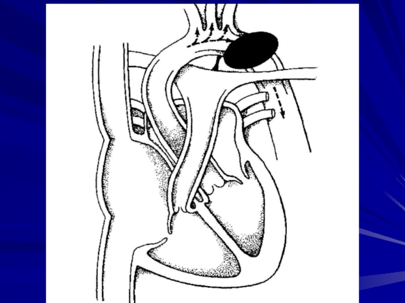
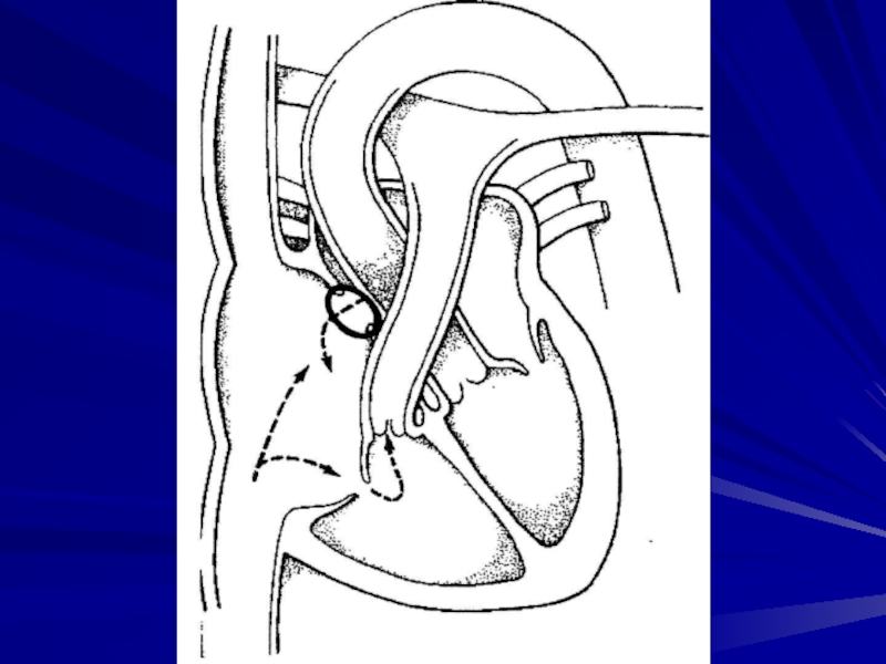
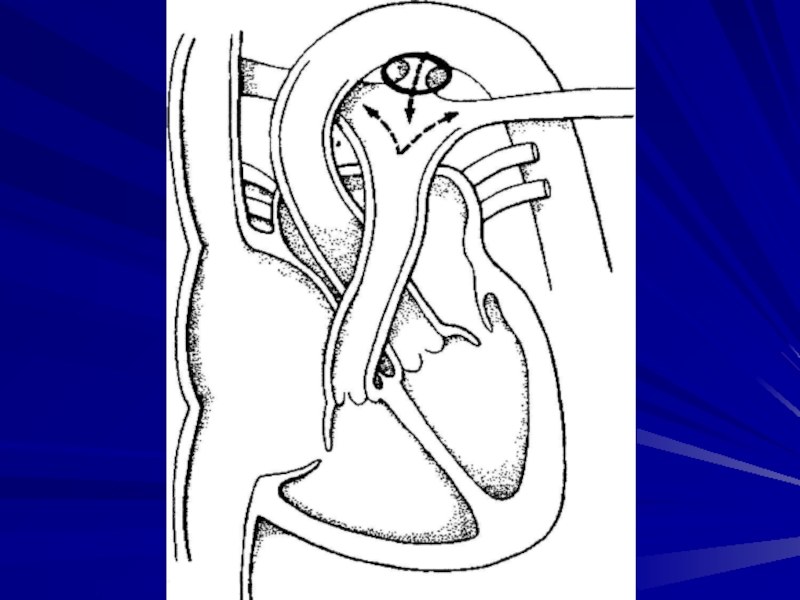
Слайд 53COARCTATION OF THE AORTA
Narrowing of the lumen results in increased systolic
blood pressure proximal to the coarctation and decreased systolic blood pressure distally. The incidence is 1:13,000 live births.