- Главная
- Разное
- Дизайн
- Бизнес и предпринимательство
- Аналитика
- Образование
- Развлечения
- Красота и здоровье
- Финансы
- Государство
- Путешествия
- Спорт
- Недвижимость
- Армия
- Графика
- Культурология
- Еда и кулинария
- Лингвистика
- Английский язык
- Астрономия
- Алгебра
- Биология
- География
- Детские презентации
- Информатика
- История
- Литература
- Маркетинг
- Математика
- Медицина
- Менеджмент
- Музыка
- МХК
- Немецкий язык
- ОБЖ
- Обществознание
- Окружающий мир
- Педагогика
- Русский язык
- Технология
- Физика
- Философия
- Химия
- Шаблоны, картинки для презентаций
- Экология
- Экономика
- Юриспруденция
Cardiac rhythm disorders in children презентация
Содержание
- 1. Cardiac rhythm disorders in children
- 2. Plan of the lecture 1. Definition
- 6. Arrhythmia reasons Cardial CHD Acquired chronic HD
- 7. Rhythm and conductivity disorders classification ( Belokon
- 8. Diagnostic approach Superficial ECG (12 traditional leads)
- 12. Normal sinus rhythm criteria Regular consecutive Р-Р
- 14. ECG criteria of sinus arrhythmia R-R
- 16. ECG criteria of sinus bradycardia QRS complexes
- 18. ECG criteria of sick sinus node syndrome
- 21. Premature Contractions (PC) can be Supraventricular or
- 22. ECG signs of premature atrium contractions (PAC)
- 24. ECG criteria of PC originated from AV-node
- 27. ECG criteria of premature ventricular contraction (PVC)
- 29. Signs of atrium paroxysmal tachycardia (PT) Wave
- 31. ECG signs of AV PT Wave
- 32. ECG signs of ventricular PT Aberrant wide
- 34. ECG signs of atrium fibrillation P-wave is
- 37. ECG signs of ventricular fibrillation QRS
- 39. ECG signs of atrium blockage Wave P
- 41. ECG signs of I grade AV blockage
- 43. Ecg signs of Mobitz-I type AV block
- 45. ECG signs of Mobitz-II AV blockage Periodic
- 46. ECG signs of III grade AV -blockage
- 48. Arrhythmias treatment Treatment of arrhythmia in children
- 49. Arrhythmias treatment Antiarrhythmic drugs are classified according
- 50. Arrhythmias treatment Beta-blockers ( propranolol-0,5 mg/kg increasing
- 51. Arrhythmias treatment Some medications that improve metabolism
- 52. Questions Prevention of cardiac rhythm

Слайд 2Plan of the lecture
1. Definition of cardiac rhythm disorders in
children
2. Etiologic factors
3. Classification
4. Clinical presentation of cardiac rhythm disorders in children
5. The differential diagnosis of cardiac rhythm disorders in children
5. Treatment
2. Etiologic factors
3. Classification
4. Clinical presentation of cardiac rhythm disorders in children
5. The differential diagnosis of cardiac rhythm disorders in children
5. Treatment


Слайд 6Arrhythmia reasons
Cardial
CHD
Acquired chronic HD
Carditis
Cardiomyopathies
Mitral valve prolapse
Cardiac neoplasms
Combined
Extracardial
Vegetative nervous system dysregulation
Endocrine disorders
CNS
diseases
Intoxications
Any somatic disease
Intoxications
Any somatic disease

Слайд 7Rhythm and conductivity disorders classification ( Belokon N.A. 1987)
1 Impulse formation
disturbance
А. Nomotope disturbance ( sinus tachycardia, bradycardia, pacemaker migration)
Б. Heterotopic rhythm disturbance (extrasystole, paroxysmal tachycardia, atrium and ventricular flutter or fibrillation)
2 Conductivity abnormalities
(sinoauricularis, ventricular, atrium, AV- blockades of 1,2, 3 grade)
3 Combined arrhythmias (sick sinus syndrome, sinus node arrest, pre-excitation syndromes, AV- dissociation)
1 Impulse formation disturbance А. Nomotope disturbance")
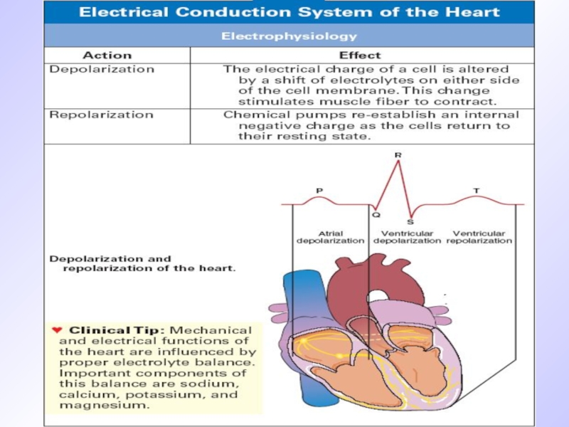
Слайд 8Diagnostic approach
Superficial ECG (12 traditional leads)
Electrophysiologic examining methods (EPM)-intracardiac or transesophageal
electrodes
HR and BP Cholter monitoring
HR and BP Cholter monitoring
Electrophysiologic examining methods (EPM)-intracardiac or transesophageal electrodes HR and BP")


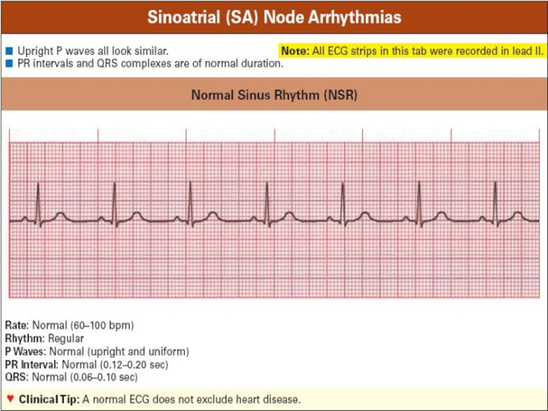
Слайд 12Normal sinus rhythm criteria
Regular consecutive Р-Р row
Constant wave P morphology
Wave P
precedes QRS complex
Normal QRS complex
Normal QRS complex

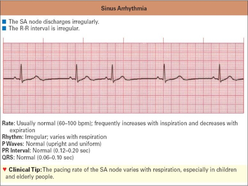
Слайд 14 ECG criteria of sinus arrhythmia
R-R interval irregular ( decreases during
inspiration)
P-P interval irregular
Wave P constantly precedes QRS complex
PR interval ranges 0,02 sec
P-P interval irregular
Wave P constantly precedes QRS complex
PR interval ranges 0,02 sec
P-P interval irregularWave P")
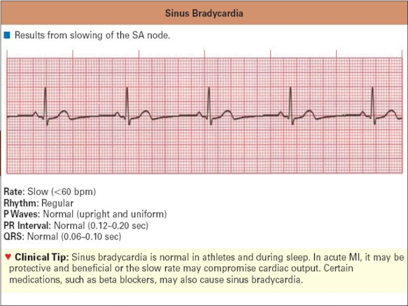
Слайд 16ECG criteria of sinus bradycardia
QRS complexes frequency less than100/min in neonates
and infants; less than 60/min in 6-9 years old children and less than 50/min. in 9-16 уears old
R-R interval is constant
Wave Р precedes every QRS complex
Interval P-R is constant not more than 0,18 sec.
R-R interval is constant
Wave Р precedes every QRS complex
Interval P-R is constant not more than 0,18 sec.

Слайд 18ECG criteria of sick sinus node syndrome
Evident tachy-brady-arrhythmia
Sinus-auricularis blockage
Atrium or/and cardiac
asystolia
When rhythm retarded less than 40/min. weakness, dizziness syncope amnesia can occur
When rhythm retarded less than 40/min. weakness, dizziness syncope amnesia can occur


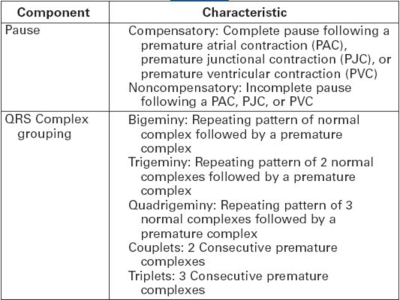
Слайд 21Premature Contractions (PC) can be
Supraventricular or ventricular
Monotopic or polytopic
Aberrant
Ultraearly, early, late
Rare,
moderate, frequent
Single, double, group
Allorhythmia
Single, double, group
Allorhythmia
 can beSupraventricular or ventricularMonotopic or polytopicAberrantUltraearly, early, lateRare, moderate, frequentSingle, double, groupAllorhythmia")
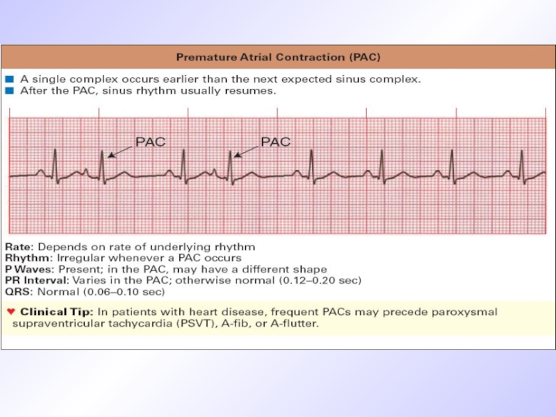
Слайд 22ECG signs of premature atrium contractions (PAC)
Short-cut preectopic interval
Wave P is
present before complex QRS
Stable shortened PQ(R)-interval
Normal narrow QRS complex, similar to previous one
Incomplete compensated pause
Stable shortened PQ(R)-interval
Normal narrow QRS complex, similar to previous one
Incomplete compensated pause
Short-cut preectopic intervalWave P is present before complex QRSStable")
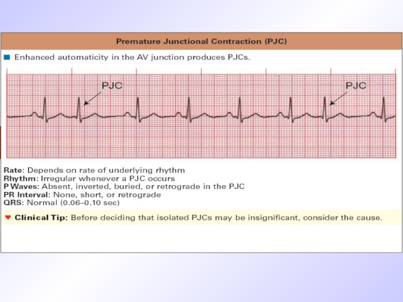
Слайд 24ECG criteria of PC originated from AV-node
Premature unstrained complex QRS
P wave
is absent before QRS
Incomplete compensated pause
Incomplete compensated pause


Слайд 27ECG criteria of premature ventricular contraction (PVC)
Wave is absent before QRS
QRS is premature aberrant, wide
ST segment is dislocated and wave T is discordant to QRS
Complete compensated pause
ST segment is dislocated and wave T is discordant to QRS
Complete compensated pause
Wave is absent before QRS QRS is premature aberrant,")

Слайд 29Signs of atrium paroxysmal tachycardia (PT)
Wave Р is present before QRS
QRS
is unstrained
HR in schoolchildren 150-160/min, in infants and toddlers– more than 200/min.
Interval PQ is relatively elongated
Segment ST is lowered, sometimes wave T is inverted
HR in schoolchildren 150-160/min, in infants and toddlers– more than 200/min.
Interval PQ is relatively elongated
Segment ST is lowered, sometimes wave T is inverted
Wave Р is present before QRSQRS is unstrainedHR in schoolchildren")

Слайд 31ECG signs of AV PT
Wave P is absent before QRS
QRS
is unstrained
HR is more than150-200/min
PQ interval is normal or elongated
Secondary changes of ST and Т
HR is more than150-200/min
PQ interval is normal or elongated
Secondary changes of ST and Т

Слайд 32ECG signs of ventricular PT
Aberrant wide regular QRS
HR 150-200/min
Constant R-R interval
Secondary
discordant segment ST and wave T changes
АV-dissociation
Reflectory maneuvres are inefficiant
АV-dissociation
Reflectory maneuvres are inefficiant


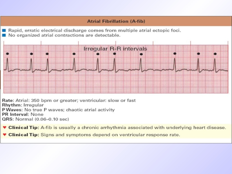
Слайд 34ECG signs of atrium fibrillation
P-wave is displaced by F-waves of different
shape and amplitude
QRS is normal but rhythm is irregular, chaotic
R-R interval changes in duration
QRS is normal but rhythm is irregular, chaotic
R-R interval changes in duration

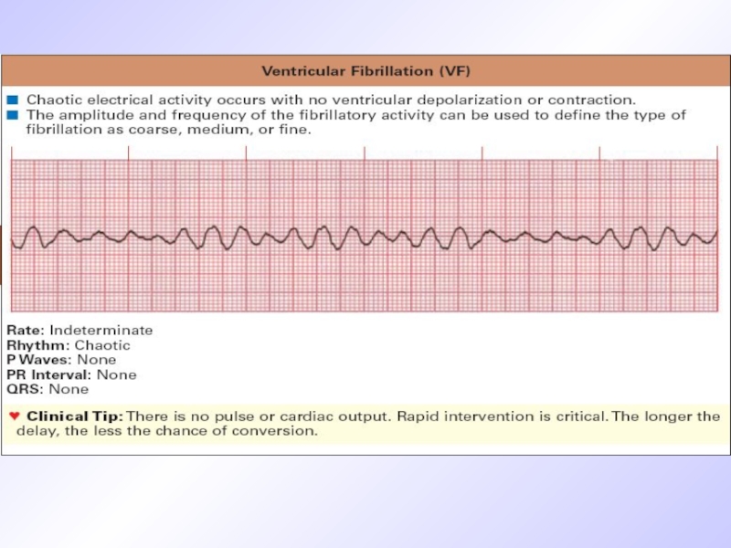
Слайд 37ECG signs of ventricular fibrillation
QRS are wide of the same
shape and amplitude
End part of QRST complex isn’t differentiated ( ST and T are absent)
Diastolic pause is absent ( isoline isn’t visualized)
Frequency of ventricular complexes is 250-300/min.
End part of QRST complex isn’t differentiated ( ST and T are absent)
Diastolic pause is absent ( isoline isn’t visualized)
Frequency of ventricular complexes is 250-300/min.

Слайд 39ECG signs of atrium blockage
Wave P is wide ( elongation to
120msec (normal one isn’t more than 95 msec)
Normal P wave amplitude
Splitting of Р wave and appearance of negative wave
PQ segment becomes shorter or disappear PR interval is normal
Normal P wave amplitude
Splitting of Р wave and appearance of negative wave
PQ segment becomes shorter or disappear PR interval is normal

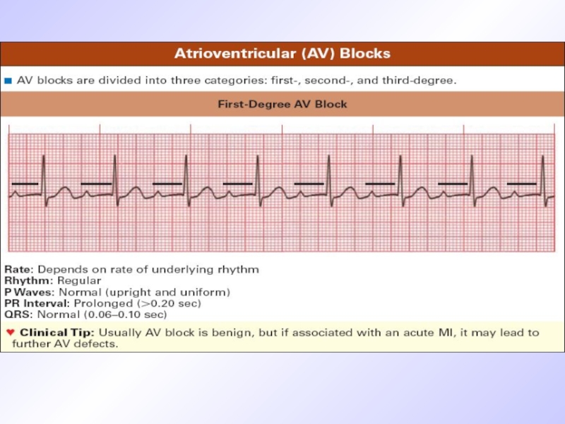
Слайд 41ECG signs of I grade AV blockage
Interval PQ elongation more than
170 ms for younger children and 200 ms for adolescents
Wave P is present after every QRS
Stable PQ interval
All QRS complexes are present
Wave P is present after every QRS
Stable PQ interval
All QRS complexes are present

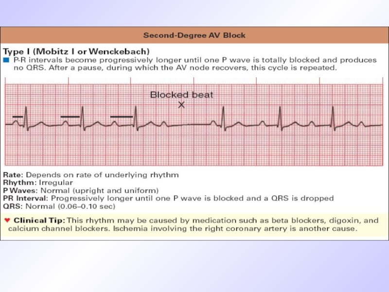
Слайд 43Ecg signs of Mobitz-I type AV block
Consecutive AV-conductivity retardation from cycle
to cycle and elongation of PQ until QRS fallout
Invariability of QRS
R-R interval before QRS missing is longer than after it.
After complex missing PQ interval restitutes again
Invariability of QRS
R-R interval before QRS missing is longer than after it.
After complex missing PQ interval restitutes again

Слайд 45ECG signs of Mobitz-II AV blockage
Periodic conductivity atrium impulse to ventricular
blockage and QRS fallout.
Stable PQ interval in all cycles
Unchangeable QRS
Regular or irregular QRS fallout with ratio of P waves to QRS as 2:1, 3:2, 4:3 etc.
Stable PQ interval in all cycles
Unchangeable QRS
Regular or irregular QRS fallout with ratio of P waves to QRS as 2:1, 3:2, 4:3 etc.

Слайд 46ECG signs of III grade AV -blockage
Complete dissociation of atrium and
ventricular contractility
P waves originate from sinus node or atrium heterotopic pacemakers
Atrium contractility frequency is according to age
Ventricular complexes are of normal morphology ( if rhythm originates from AV node ) or aberrant if rhythm is ideoventricular
Ventricular rhythm is1,5-2 times less than atrium one ( 40-65/min)
Different rhythm rate and dissociation of atrium and ventricular contractility lead to chaotic P wave location as for QRS.


Слайд 48Arrhythmias treatment
Treatment of arrhythmia in children differs from therapy in adults.
Main approach is to treat reasons that cause development of rhythm disorders (i.e. inflammatory processes, endocrine diseases, vegetative or metabolic disorders). Only in cases of threatening to life arrhythmias anti-arrhythmic drugs can be used

Слайд 49Arrhythmias treatment
Antiarrhythmic drugs are classified according E. Vaughan-Williams (1984) for IV
classes
Class I membrane stabilizers (lidocain)
Class II Beta-blockers (propranolol)
Class III medications that prolong repolarization phase (amiodaron)
Class IV –Ca-channels blockers (verapamil, diltiazem)
Class I membrane stabilizers (lidocain)
Class II Beta-blockers (propranolol)
Class III medications that prolong repolarization phase (amiodaron)
Class IV –Ca-channels blockers (verapamil, diltiazem)
 for IV classesClass I membrane stabilizers")
Слайд 50Arrhythmias treatment
Beta-blockers ( propranolol-0,5 mg/kg increasing dosage to 3-5 mg/kg/day steadily,
atenolol 1-2 mg/kg bid, nadolol 1-3 mg/kg/day)- in supraventricular tachycardias or premature beats, sometimes in ventricular ones
Amiodaron or cordaron (5-15 mg/kg/day bid 2 weks, then steadily dosage must be decreased)-is effective in both supraventricular and ventricular rhythm disorders
Lidocain (0,5-1 mg/kg for first 2 hours, then 1-2 mg/min IV slowly) – only for ventricular tachycardia, premature beats
Amiodaron or cordaron (5-15 mg/kg/day bid 2 weks, then steadily dosage must be decreased)-is effective in both supraventricular and ventricular rhythm disorders
Lidocain (0,5-1 mg/kg for first 2 hours, then 1-2 mg/min IV slowly) – only for ventricular tachycardia, premature beats

Слайд 51Arrhythmias treatment
Some medications that improve metabolism of cardiomyocytes has also indirect
anti-arrhythmic activity
mildronat,
L-carnitin,
preductal,
Magne-B6, magnerot
Riboxyn,
panangyn or asparcam,
vitamins - antioxydants like triovit, vitamax
mildronat,
L-carnitin,
preductal,
Magne-B6, magnerot
Riboxyn,
panangyn or asparcam,
vitamins - antioxydants like triovit, vitamax

Слайд 52
Questions
Prevention of cardiac rhythm disorders in children
Frequency and prognosis
Common clinical
symptoms of cardiac rhythm disorders in children
Additional (instrumental) methods of invastigations
Prevention of complications.
Principles of treatment of cardiac rhythm disorders in children
Additional (instrumental) methods of invastigations
Prevention of complications.
Principles of treatment of cardiac rhythm disorders in children