- Главная
- Разное
- Дизайн
- Бизнес и предпринимательство
- Аналитика
- Образование
- Развлечения
- Красота и здоровье
- Финансы
- Государство
- Путешествия
- Спорт
- Недвижимость
- Армия
- Графика
- Культурология
- Еда и кулинария
- Лингвистика
- Английский язык
- Астрономия
- Алгебра
- Биология
- География
- Детские презентации
- Информатика
- История
- Литература
- Маркетинг
- Математика
- Медицина
- Менеджмент
- Музыка
- МХК
- Немецкий язык
- ОБЖ
- Обществознание
- Окружающий мир
- Педагогика
- Русский язык
- Технология
- Физика
- Философия
- Химия
- Шаблоны, картинки для презентаций
- Экология
- Экономика
- Юриспруденция
Bullous and vesicular dermatoses презентация
Содержание
- 1. Bullous and vesicular dermatoses
- 2. Theoretical part The group of vesicular and
- 3. True (acantholytic) pemphigus Pemphigus is a malignant,
- 4. Etiology and pathogenesis of pemphigus There are
- 5. Clinical varieties Four forms of true
- 6. Pemphigus Vulgaris This form of pemphigus
- 7. Pemphigus Vulgaris Usually dermatosis begins with affection
- 8. Pemphigus Vegetans At the beginning of
- 9. Pemphigus Vegetans Papulomatous growths secreting a considerable
- 10. Pemphigus Foliaceus The disease is characterized
- 11. Pemphigus seborrhoicus, or erythematosus (Senear-Usher syndrome) Pemphigus
- 12. Pemphigus seborrhoicus, or erythematosus The lesions usually
- 13. Diagnosis and differential diagnosis Differential diagnosis is
- 14. Dermatitis herpetiformis (Duhring’s disease)
- 15. Herpes simplex The disease is caused
- 16. Treatment Treatment with steroid hormones envisages the
- 17. Treatment. Drying and disinfectant agents are
- 18. The following clinical forms are distinguished:
- 19. Histopathology The characteristic findings are ballooning
- 20. Herpes zoster Herpes zoster, also called
- 21. The following clinical varieties are distinguished:
- 22. Histopathology. Ballooning and reticular degeneration of
- 23. Treatment. Antiviral drugs such as methisazonum
Слайд 2Theoretical part
The group of vesicular and bullosus dermatoses includes different diseases
on the basis of etiology and pathogenesis (pemphigus, Duhring’s dermatosis, simple vesicular lichens, herpes zoster, exudative multimorphic erythema).
Слайд 3True (acantholytic) pemphigus
Pemphigus is a malignant, serious disease. Its clinical manifestation
is the formation of vesicles on non-inflamed skin and mucous membranes. If not treated the bulloses soon appear on the whole skin. Patients should consult not only dermatologists but also other specialists (physicians, dentists, infectionists). Due to this, the knowledge of this pathology is necessary for all the clinicians to render qualified help to the patients.
Слайд 4Etiology and pathogenesis of pemphigus
There are different etiopathogenic theories, in particular,
the viral theory but it is not completely proved. Recently autoimmune processes are considered to be of great importance in the pathogenesis: discovery of antibodies to intercellular substance in the skin, in liquid of the bulloses and in blood serum. In immunofluorescence, in intercellular space of stratum spinosum of epidermis immunoglobulin G is found only in the patients with pemphigus.
Слайд 5Clinical varieties
Four forms of true pemphigus are differentiated:
pemphigus vulgaris
(common) pemphigus vegetans
pemphigus foliaceus (exfoliative) seborrheal pemphigus
pemphigus foliaceus (exfoliative) seborrheal pemphigus
Слайд 6Pemphigus Vulgaris
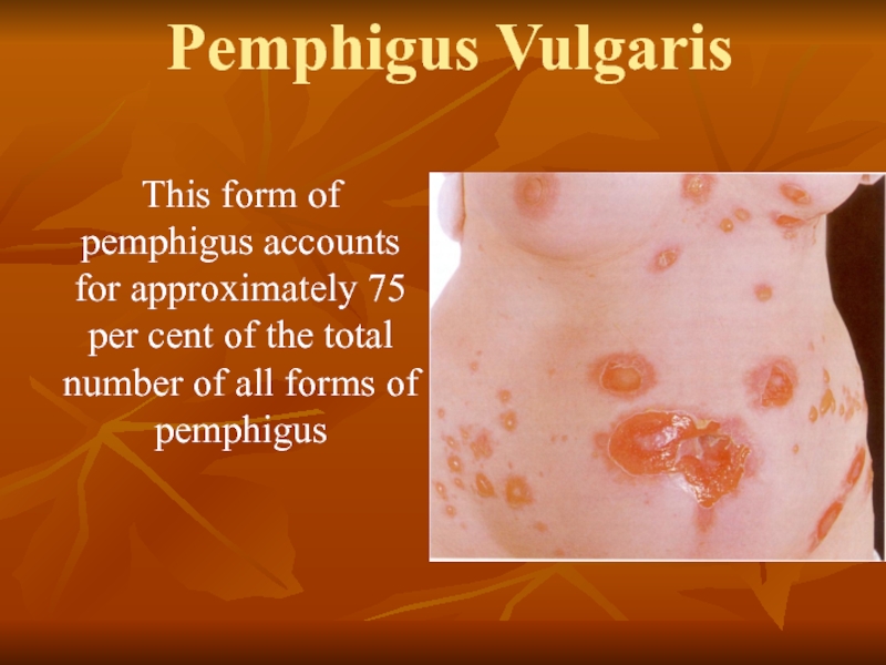
This form of pemphigus accounts for approximately 75 per cent
of the total number of all forms of pemphigus
Слайд 7Pemphigus Vulgaris
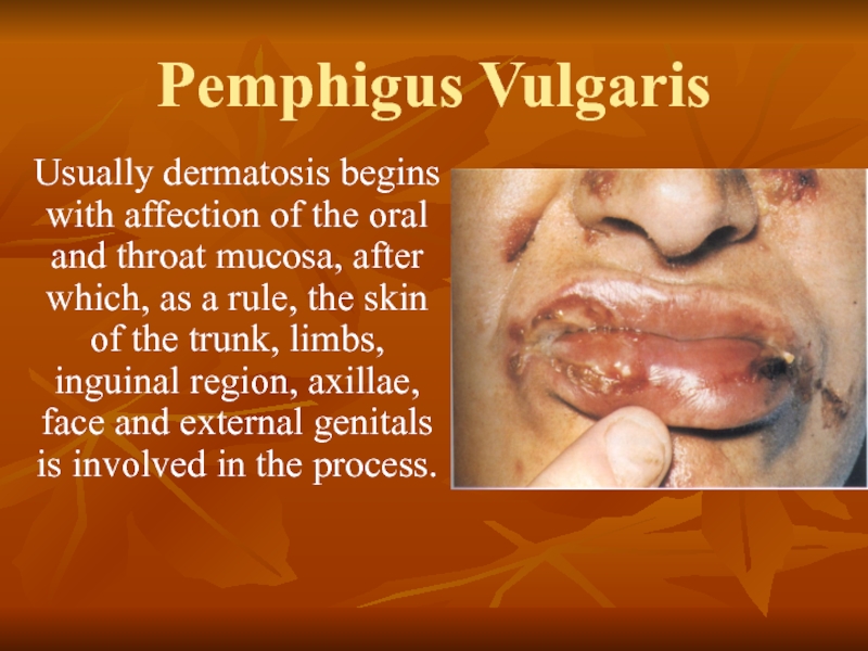
Usually dermatosis begins with affection of the oral and throat
mucosa, after which, as a rule, the skin of the trunk, limbs, inguinal region, axillae, face and external genitals is involved in the process.
Слайд 8Pemphigus Vegetans
At the beginning of its development this form of pemphigus
is clinically similar to pemphigus vulgaris and often starts with the appearance of lesions on the oral mucosa. From the very onset of the disease, however, attention is drawn to the tendency of the bullae to be localized around the natural orifices, the navel and in the region of the large skin folds
Слайд 9Pemphigus Vegetans
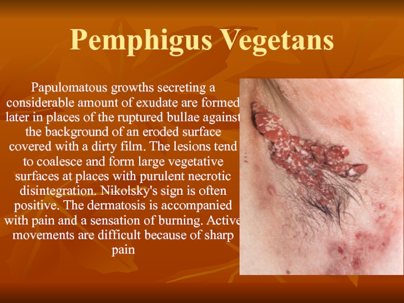
Papulomatous growths secreting a considerable amount of exudate are formed
later in places of the ruptured bullae against the background of an eroded surface covered with a dirty film. The lesions tend to coalesce and form large vegetative surfaces at places with purulent necrotic disintegration. Nikolsky's sign is often positive. The dermatosis is accompanied with pain and a sensation of burning. Active movements are difficult because of sharp pain
Слайд 10Pemphigus Foliaceus
The disease is characterized by drastic acantholysis leading to the
formation of superficial fissures directly under the horny layer, which later turn into bullae.
At the beginning of the disease, flaccid bullae with a thin top and slightly elevated above the surface form on apparently healthy skin. They rupture rapidly with the formation of large erosions. More frequently the tops of the bullae dry up into thin stratified scaly crusts. Epithelization of erosions under the crusts is slow. New portions of the exudate cause the layering of these crusts, producing a scaly surface, hence there is a term 'exfoliative', by which the disease is also known. It is in this variant of pemphigus that the sign described by Nikolsky in 1896 is always sharply positive.
At the beginning of the disease, flaccid bullae with a thin top and slightly elevated above the surface form on apparently healthy skin. They rupture rapidly with the formation of large erosions. More frequently the tops of the bullae dry up into thin stratified scaly crusts. Epithelization of erosions under the crusts is slow. New portions of the exudate cause the layering of these crusts, producing a scaly surface, hence there is a term 'exfoliative', by which the disease is also known. It is in this variant of pemphigus that the sign described by Nikolsky in 1896 is always sharply positive.
Слайд 11Pemphigus seborrhoicus,
or erythematosus (Senear-Usher syndrome)
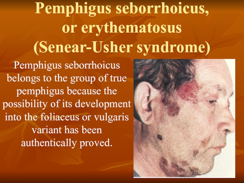
Pemphigus seborrhoicus belongs to the group of
true pemphigus because the possibility of its development into the foliaceus or vulgaris variant has been authentically proved.
Слайд 12Pemphigus seborrhoicus,
or erythematosus
The lesions usually first appear on the face and
less frequently on the scalp, chest and back. Closely arranged greasy crusts are formed against an erythematous background and simulate the picture of cicatrizing erythematosis on the face. Moist eroded surfaces are exposed when the crusts are removed. Acantholytic cells are seen in impression smears from these surfaces. The bullae are often formed unnoticeably and the crusts seem to be primary lesions. In other cases vesicles covered with stratified yellowish crusts are formed on the trunk and limbs, in sites characteristic of seborrhea. Lesions rarely occur on the mucous membranes, but if this happens, they are a bad prognostic sign.
Слайд 13Diagnosis and differential diagnosis
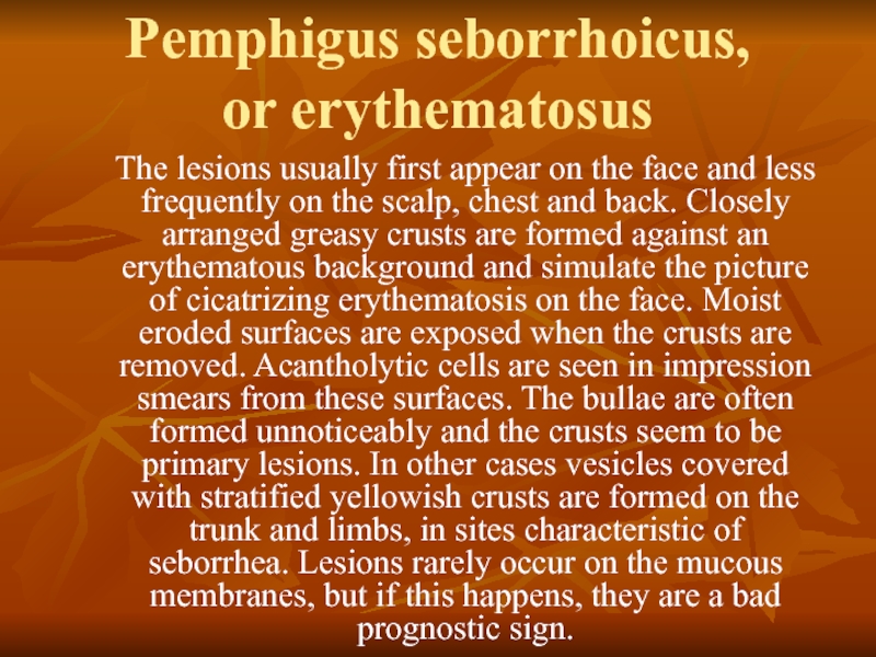
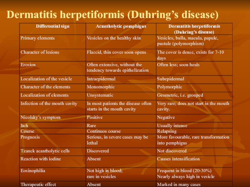
Differential diagnosis is carried out with multiform exudative
erythema, bullous toxicoderma (Lyell’s syndrome) and Duhring’s dermatosis
Слайд 15Herpes simplex
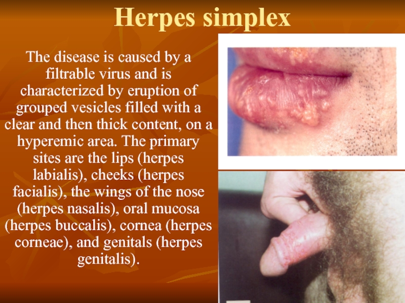
The disease is caused by a filtrable virus and is
characterized by eruption of grouped vesicles filled with a clear and then thick content, on a hyperemic area. The primary sites are the lips (herpes labialis), cheeks (herpes facialis), the wings of the nose (herpes nasalis), oral mucosa (herpes buccalis), cornea (herpes corneae), and genitals (herpes genitalis).
Слайд 16Treatment
Treatment with steroid hormones envisages the prescription of maximum doses (80-100
mg of prednisolone or methylprednisolone, 4‑6 mg of dexamethasone daily). When new eruptions cease to appear and exudative phenomena in the foci diminishes, the dose of corticosteroids may be reduced, but very slowly (to avoid exacerbation of the process and prevent the 'phenomenon of steroid drug discontinuation').
Слайд 17Treatment.
Drying and disinfectant agents are used externally: lotions of Aq.
Goulardi and 1-4 per cent silver nitrate solution, 1-2 per cent pyoktanin (gentian violet) solution, 1-2-3 per cent oxolinum or 30-50 per cent interferon ointment, Bonaphton, gossypol, tebrophenum, 1-3 per cent Florenal ointment, and sulfur-carbolic paste (Ac. carbolici 1.0, Sulfuris 1.5, Pastae Zinci 30.0).
Слайд 18The following clinical forms are distinguished:
mild; with rapid resolution of
the few lesions that have erupted;
edematous; accompanied with bright hyperemia and marked swelling;
severe;
zosteriform;
frequently recurring; localized on the lips, buttocks, and external genitals.
edematous; accompanied with bright hyperemia and marked swelling;
severe;
zosteriform;
frequently recurring; localized on the lips, buttocks, and external genitals.
Слайд 19Histopathology
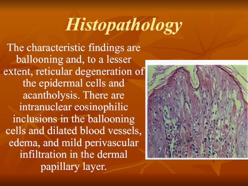
The characteristic findings are ballooning and, to a lesser extent,
reticular degeneration of the epidermal cells and acantholysis. There are intranuclear eosinophilic inclusions in the ballooning cells and dilated blood vessels, edema, and mild perivascular infiltration in the dermal papillary layer.
Слайд 20Herpes zoster
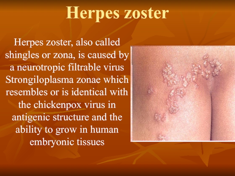
Herpes zoster, also called shingles or zona, is caused by
a neurotropic filtrable virus Strongiloplasma zonae which resembles or is identical with the chickenpox virus in antigenic structure and the ability to grow in human embryonic tissues
Слайд 21The following clinical varieties are distinguished:
generalized herpes zoster marked by
bilateral and disseminated lesions;
herpes zoster haemorrhagicus, in which the clear contents of the vesicles turn purulent and then, when the process penetrates deeper into the dermis, become hemorrhagic;
herpes zoster gangraenosus, a severe form, in which the floor of the vesicles undergoes necrosis and scars are formed in their place;
mild form;
bullous form characterized by the appearance of both vesicles and bullae.
Слайд 22Histopathology.
Ballooning and reticular degeneration of the epidermal cells, intranuclear viral
inclusions, and degenerative changes in the nerve fibres may be seen. Acute inflammatory polymorphonuclear infiltration predominantly of a lymphocyto-histio-cytic character, edema, and dilated blood and lymph vessels are revealed.
Слайд 23Treatment.
Antiviral drugs such as methisazonum or cutizonum (one taken two
or three times a day for three to six days), interferon, salicylates and analgesics (acetyl salicylic acid, amidopyrine, phenylbutazone, rheopyrine), vitamins B1, B5 B6, B12, and C, autohaemotherapy, gamma globulin injections, and interferonogens are prescribed.
