- Главная
- Разное
- Дизайн
- Бизнес и предпринимательство
- Аналитика
- Образование
- Развлечения
- Красота и здоровье
- Финансы
- Государство
- Путешествия
- Спорт
- Недвижимость
- Армия
- Графика
- Культурология
- Еда и кулинария
- Лингвистика
- Английский язык
- Астрономия
- Алгебра
- Биология
- География
- Детские презентации
- Информатика
- История
- Литература
- Маркетинг
- Математика
- Медицина
- Менеджмент
- Музыка
- МХК
- Немецкий язык
- ОБЖ
- Обществознание
- Окружающий мир
- Педагогика
- Русский язык
- Технология
- Физика
- Философия
- Химия
- Шаблоны, картинки для презентаций
- Экология
- Экономика
- Юриспруденция
Асфиксия и рН пуповинной крови презентация
Содержание
- 1. Асфиксия и рН пуповинной крови
- 3. Критерии асфиксии 1996 The American Academy of
- 4. Критерии асфиксии The second Consensus statement was
- 5. Критерии асфиксии The third consensus statement was
- 6. The cornerstone of all three statements
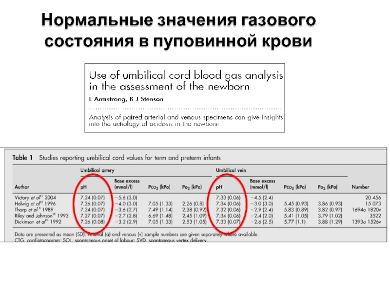
- 7. Нормальные значения газового состояния в пуповинной крови
- 8. У каждого второго ребенка рН меньше 7,0, почему этот ребенок умер?
- 9. Goodwin TM, Belai I, Hernandez P, et
- 10. Анализ газового состояния пуповинной крови рекомендуется проводит
- 11. Анализ газового состояния пуповинной крови ребенка рН
- 12. Выводы: Отрицательные моменты: В данном случае, на
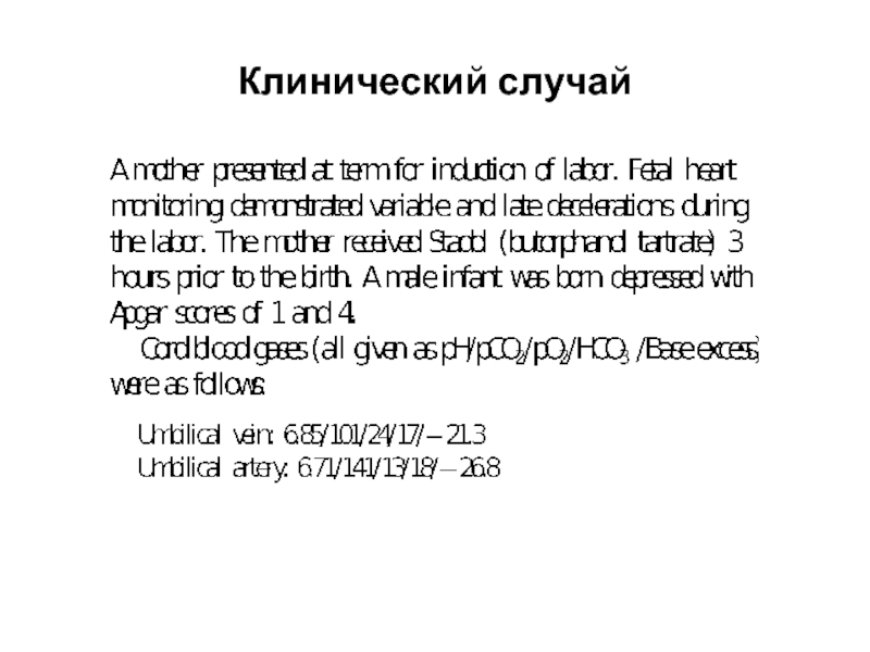
- 13. Клинический случай
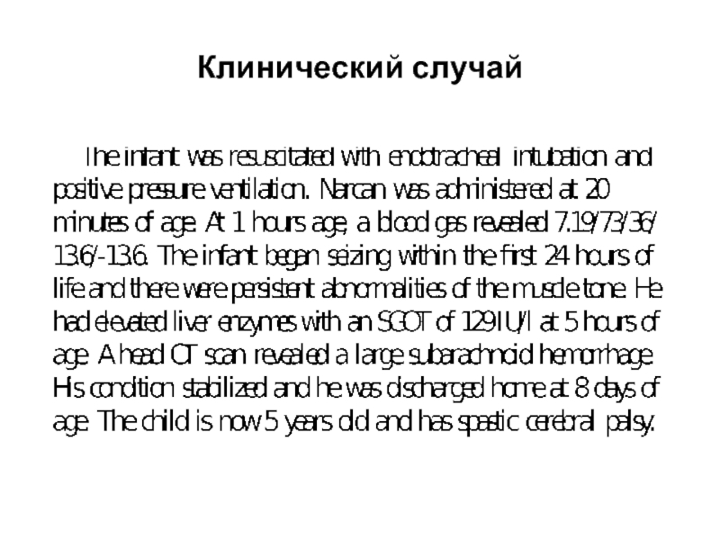
- 14. Клинический случай
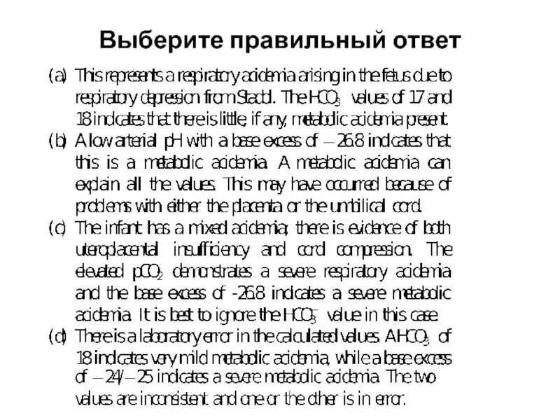
- 15. Выберите правильный ответ
- 16. Помогает ли электронный мониторинг плода предупредит ацидоз у плода?
- 17. Electronic Fetal Monitoring Jennifer Merriman, MD Delaware Center for Maternal & Fetal Medicine
- 18. Randomized Trials regarding EFM – Part 1
- 19. Can EFM predict fetal acidemia? Many studies
- 20. Can EFM predict fetal acidemia? HOWEVER …
- 21. Назарларыңызға көп рахмет!
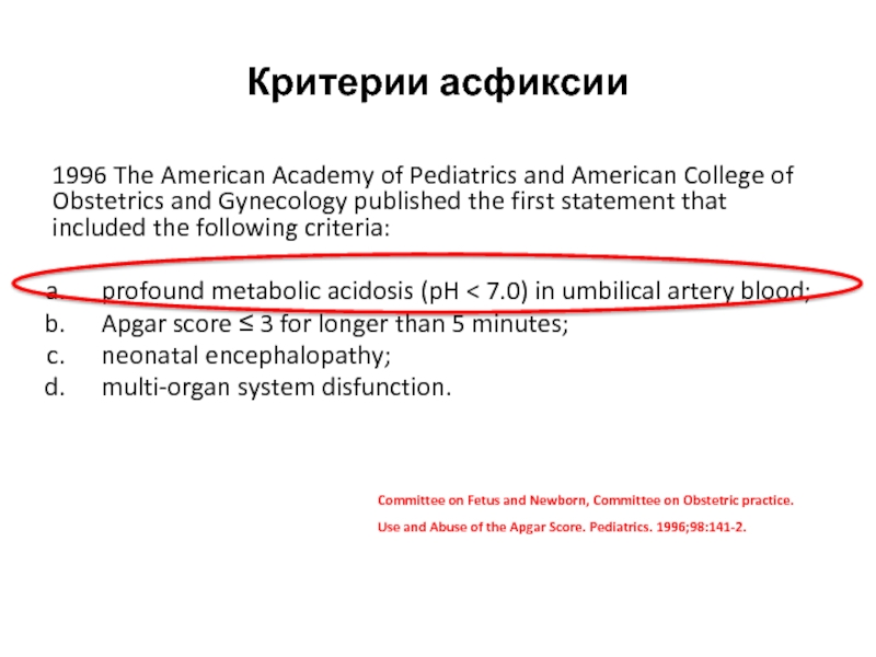
Слайд 3Критерии асфиксии
1996 The American Academy of Pediatrics and American College of
profound metabolic acidosis (pH < 7.0) in umbilical artery blood;
Apgar score ≤ 3 for longer than 5 minutes;
neonatal encephalopathy;
multi-organ system disfunction.
Committee on Fetus and Newborn, Committee on Obstetric practice. Use and Abuse of the Apgar Score. Pediatrics. 1996;98:141-2.
Слайд 4Критерии асфиксии
The second Consensus statement was approved by the International Cerebral
The essential criteria were the following:
metabolic acidosis in early neonatal blood sample (pH < 7.0 and base deficit ≥ 12 mmol/L);
moderate or severe encephalopathy;
cerebral palsy of spastic quadriplegia, dyskinetic or mixed type.
The 5 additional criteria were:
sentinel event;
severe changes in fetal heart rate;
Apgar score < 6 beyond 5 min;
multi-system involvement;
early imaging evidence [6].
MacLennan A. A template for defining a causal relation between
acute intrapartum events and CP: International Consensus Statement.
BMJ. 1999;319(7216):1054-9.
Слайд 5Критерии асфиксии
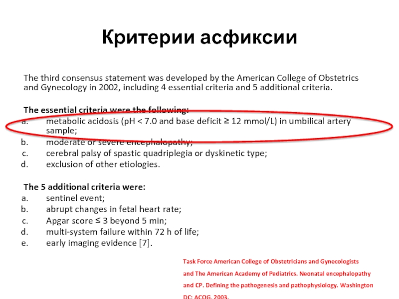
The third consensus statement was developed by the American College
The essential criteria were the following:
metabolic acidosis (pH < 7.0 and base deficit ≥ 12 mmol/L) in umbilical artery sample;
moderate or severe encephalopathy;
cerebral palsy of spastic quadriplegia or dyskinetic type;
exclusion of other etiologies.
The 5 additional criteria were:
sentinel event;
abrupt changes in fetal heart rate;
Apgar score ≤ 3 beyond 5 min;
multi-system failure within 72 h of life;
early imaging evidence [7].
Task Force American College of Obstetricians and Gynecologists
and The American Academy of Pediatrics. Neonatal encephalopathy and CP. Defining the pathogenesis and pathophysiology. Washington DC: ACOG, 2003.
Слайд 6
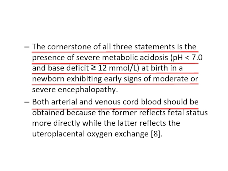
The cornerstone of all three statements is the presence of severe
Both arterial and venous cord blood should be obtained because the former reflects fetal status more directly while the latter reflects the uteroplacental oxygen exchange [8].
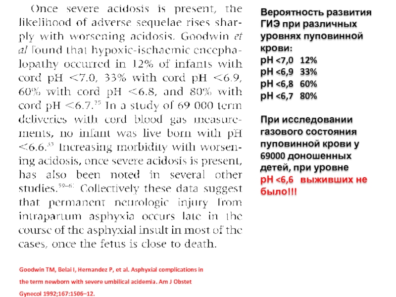
Слайд 9Goodwin TM, Belai I, Hernandez P, et al. Asphyxial complications in
Вероятность развития ГИЭ при различных уровнях пуповинной крови:
рН <7,0 12%
рН <6,9 33%
рН <6,8 60%
рН <6,7 80%
При исследовании газового состояния пуповинной крови у 69000 доношенных детей, при уровне
рН <6,6 выживших не было!!!
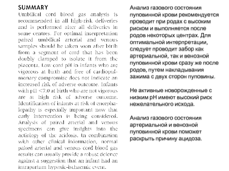
Слайд 10Анализ газового состояния пуповинной крови рекомендуется проводит при родах с высоким
Не активные новорожденные с низким рН имеют высокий риск нежелательного исхода.
Анализ газового состояния артериальной и венозной пуповинной крови поможет раскрыть причину ацидоза.
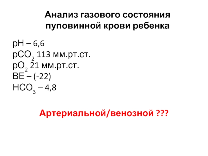
Слайд 11Анализ газового состояния пуповинной крови ребенка
рН – 6,6
рСО2 113 мм.рт.ст.
рО2 21
Артериальной/венозной ???
Слайд 12Выводы:
Отрицательные моменты:
В данном случае, на неонатальном этапе, вероятность предотвращения смерти была
Положительные моменты:
В центре проводится анализ газового состояния пуповинной крови
Предложения:
Необходимо проводить забор артериальной и венозной пуповинной крови для анализа газового состояния
Слайд 17Electronic Fetal Monitoring
Jennifer Merriman, MD
Delaware Center for Maternal & Fetal Medicine
Слайд 18Randomized Trials regarding EFM – Part 1
1986 – Leveno publication in
34,995 pregnancies assessed
No difference in rate of stillbirth, low Apgar scores, need for fetal ventilation, fetal seizures or NICU admission; however there was an increase in cesarean delivery for fetal distress
Follow-up studies including one by Vinzileos in 1993 randomized patients to continuous fetal monitoring versus intermittent fetal monitoring
No difference in Apgar scores, fetal acidosis at birth, neonatal resuscitation or other neonatal complications
Increased rate of cesarean delivery and assisted vaginal birth
2006 – Cochrane metanalysis comparing continuous EFM vs. intermittent EFM
Increased risk of cesarean delivery (RR 1.66; 95% confidence interval [CI], 1.30–2.13)
Increased the risk of Vacuum and forceps operative vaginal delivery (RR, 1.16; 95% CI, 1.01–1.32).
No reduction in perinatal mortality (RR, 0.85; 95% CI, 0.59–1.23).
Reduced risk of neonatal seizures (RR, 0.50; 95% CI, 0.31–0.80).
No reduction in the risk of cerebral palsy (RR, 1.74; 95% CI, 0.97–3.11).
Слайд 19Can EFM predict fetal acidemia?
Many studies have evaluated this question in
Various components of EFM during labor such as presence of decelerations, fetal tachycardia, reduced variability and/or fetal bradycardia have been evaluated for a possible link with neonatal encephalopathy
Studies from 2003-2007 note that there may be an association between EFM patterns and umbilical artery pH; however there are no clear EFM patterns specifically associated with fetal acidemia
Слайд 20Can EFM predict fetal acidemia?
HOWEVER … fetal variability and/or fetal accelerations
THEREFORE, in a fetus with moderate variability and/or fetal accelerations on EFM, hypoxic-induced metabolic ischemia can be reliably excluded
To date, NO evidence proving that electronic EFM reduces the rate of neonatal encephalopathy!
No evidence in the current literature that supports the ability of practitioners to predict neonatal neurologic injury, cerebral palsy or stillbirth using EFM.
Neonatal Encephalopathy & Neurologic Outcome, 2nd Edition, 2014

