- Главная
- Разное
- Дизайн
- Бизнес и предпринимательство
- Аналитика
- Образование
- Развлечения
- Красота и здоровье
- Финансы
- Государство
- Путешествия
- Спорт
- Недвижимость
- Армия
- Графика
- Культурология
- Еда и кулинария
- Лингвистика
- Английский язык
- Астрономия
- Алгебра
- Биология
- География
- Детские презентации
- Информатика
- История
- Литература
- Маркетинг
- Математика
- Медицина
- Менеджмент
- Музыка
- МХК
- Немецкий язык
- ОБЖ
- Обществознание
- Окружающий мир
- Педагогика
- Русский язык
- Технология
- Физика
- Философия
- Химия
- Шаблоны, картинки для презентаций
- Экология
- Экономика
- Юриспруденция
Microscope Measurement презентация
Содержание
- 1. Microscope Measurement
- 2. Microscope Measurement How big is that object in the microscope?
- 3. Lesson Objectives Calculate the magnification using different
- 6. Light Microscope A light microscope (also, optical microscope) is
- 7. Electron Microscope An electron microscope is an optical
- 9. Light microscope vs Electron microscope
- 10. What is happening to the image as you increase the power of the objective lens?
- 11. Calculating total magnification If two lenses
- 12. How do we find the overall magnification
- 13. 1000 1000 1000 1000 mm Micrometre nm
- 14. 0.001 0.000001 1000 0.001 1 000 000 1000 3000 0.003 0.007 0.000007 500 000 500
- 16. The diagram below is a drawing of
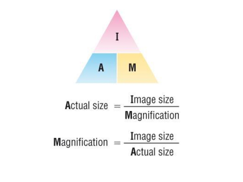
- 17. Calculating actual size: Size of the magnified image > actual size
- 18. To accurately measure the size of cellular structures we need a suitable scale:
- 19. Field of View When you look into
- 20. Estimating Specimen Size The area of the
- 21. Ideally, we need a scale we can
- 22. Eye piece graticule or reticule It is
- 23. Stage Micrometer simply a microscope slide with
- 24. Instructions Take sample of onion cell (peel
- 25. IMPORTANT FORMULA! Object Size = field of


Слайд 3Lesson Objectives
Calculate the magnification using different objective lens.
Differentiate between eyepiece graticule
and the stage micrometer.
Convert mm to micrometers.
Calculate the cell length and breadth using the relationship between the size of the image, actual size and magnification.
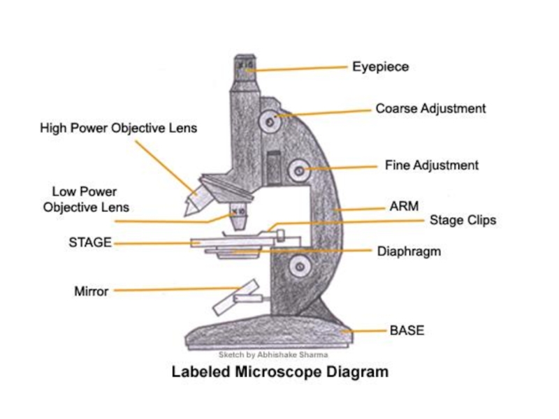
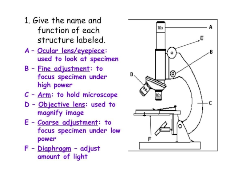
The structure and function of different parts of the microscope
The difference between a light microscope and an electron microscope.
Convert mm to micrometers.
Calculate the cell length and breadth using the relationship between the size of the image, actual size and magnification.
The structure and function of different parts of the microscope
The difference between a light microscope and an electron microscope.

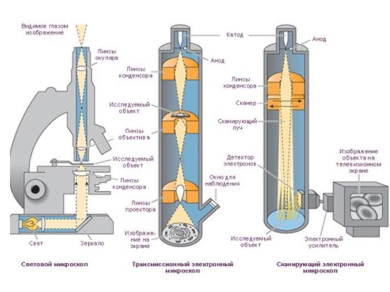
Слайд 6Light Microscope
A light microscope (also, optical microscope) is an optical instrument used to
make objects larger in order to view their details. It uses light to illuminate the objects under view
 is an optical instrument used to make objects larger in")
Слайд 7Electron Microscope
An electron microscope is an optical instrument that uses a beam
of electrons to make objects larger for a detailed view

Слайд 9
Light microscope vs Electron microscope
What is the difference between a light
microscope and an electron microscope? A number of differences such as the source of light they use, their magnification level, cost, resolving power, among other factors sets these two types of microscopes apart from each other.
VIDEO
VIDEO


Слайд 11Calculating total magnification
If two lenses are always magnifying the specimen, how
do you figure out the total magnification being used ?
Total Magnification = ocular x objective
= 10 x 4 (low power)
= 40 (low power)
Total Magnification = ocular x objective
= 10 x 4 (low power)
= 40 (low power)

Слайд 12How do we find the overall magnification of a light microscope?
Eyepiece
Objective
lens
40
100
400
1000


Слайд 16The diagram below is a drawing of an organelle from a
ciliated cell as seen with an electron microscope.
Calculate the actual length of the organelle as shown by the line AB in the diagram. Express your answer to the nearest micrometer (mm).
Show your working.
Answer = ........................................... μm
Calculate the actual length of the organelle as shown by the line AB in the diagram. Express your answer to the nearest micrometer (mm).
Show your working.
Answer = ........................................... μm
A =
I
M
=
102mm
20000
=
102000μm
20000
5.1


Слайд 19Field of View
When you look into a microscope, the “field of
view” is the visible circular area.
What happens to your field of view when you increase the power of the objective lens?
By knowing the size of the field of view (diameter), you can measure the size of objects in the microscope.
The size of objects in the field of view is different at each magnification you have to calculate the diameters of the fields of view at each magnification.
This process is called “calibrating your microscope”

Слайд 20Estimating Specimen Size
The area of the slide that you see when
you look through a microscope is called the "Field of View".
If you know how wide your field of view is, you can estimate the size of things you see in the field of view.
If you know how wide your field of view is, you can estimate the size of things you see in the field of view.


Слайд 22Eye piece graticule or reticule
It is a glass or plactic disc
with 8 divisions etched on to its surface and fitted into one eyepiece.
The size of the eyepiece reticule is constant despite the change in magnification of the object.
The value of each division varies with the change in magnification.
The size of the eyepiece reticule is constant despite the change in magnification of the object.
The value of each division varies with the change in magnification.

Слайд 23Stage Micrometer
simply a microscope slide with a finely divided scale marked
on the surface.
1 division= 0.01 mm
10 divisions= 0.1 mm
100 divisions = 1 mm
1 mm = 1000 micrometers.
1 division= 0.01 mm
10 divisions= 0.1 mm
100 divisions = 1 mm
1 mm = 1000 micrometers.

Слайд 24Instructions
Take sample of onion cell (peel of the onion)
Add a drop
of water
Cover the subject glass with cover slip
Fix it with the stage clips.
Focus the specimen on low power objective lens.
Now change to medium power objective lens and observe.
Change to high power objective lens and observe.
Cover the subject glass with cover slip
Fix it with the stage clips.
Focus the specimen on low power objective lens.
Now change to medium power objective lens and observe.
Change to high power objective lens and observe.
Add a drop of waterCover the subject")
Слайд 25IMPORTANT FORMULA!
Object Size = field of view (in mm) • 1000
number of “fits”
Object Size = ? µm
** Remember that the field of view changes with each objective!.
 • 1000")