- Главная
- Разное
- Дизайн
- Бизнес и предпринимательство
- Аналитика
- Образование
- Развлечения
- Красота и здоровье
- Финансы
- Государство
- Путешествия
- Спорт
- Недвижимость
- Армия
- Графика
- Культурология
- Еда и кулинария
- Лингвистика
- Английский язык
- Астрономия
- Алгебра
- Биология
- География
- Детские презентации
- Информатика
- История
- Литература
- Маркетинг
- Математика
- Медицина
- Менеджмент
- Музыка
- МХК
- Немецкий язык
- ОБЖ
- Обществознание
- Окружающий мир
- Педагогика
- Русский язык
- Технология
- Физика
- Философия
- Химия
- Шаблоны, картинки для презентаций
- Экология
- Экономика
- Юриспруденция
What is infection презентация
Содержание
- 1. What is infection
- 2. WHAT IS INFECTION? Infection is the invasion of
- 3. WHAT CAUSES INFECTIONS? Infections are caused by infectious agents including Bacteria Viruses Fungi (ringworm) Protozoa Macroparasites (nematodes, tapeworms, helminths)
- 5. PROPERTIES OF INFECTIOUS AGENTS Pathogenicity Virulence Specificity Organotropicity
- 6. PATHOGENICITY Pathogenicity is a specific sign of
- 7. VIRULENCE Virulence is the degree of pathogenicity
- 8. SPECIFICITY Each infectious disease causes a specific
- 9. ORGANOTROPICITY Organotropicity is the defeat of cells,
- 10. VIRULENCE FACTORS 1. Adhesion to cells Many
- 11. VIRULENCE FACTORS 2. Invasiveness Some virulent bacteria
- 12. VIRULENCE FACTORS 3. Colonization is the process
- 13. VIRULENCE FACTORS 4. Suppression of the immune
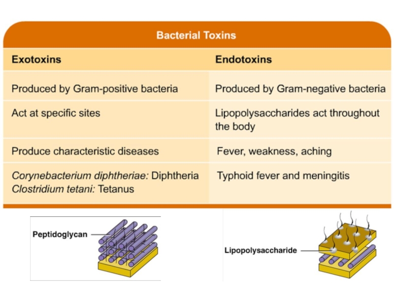
- 14. VIRULENCE FACTORS 5. Toxins Many virulence factors
- 15. BACTERIAL TOXINS
- 18. FORMS OF INFECTION PROCESSES Infectious disease
- 19. COMMON ORAL INFECTIONS Oral Herpes Gingivitis Periodontal Disease Dental Caries Canker Sores
- 20. REASONS OF ORAL INFECTIONS injuries a lack
- 21. BIOLOGICAL RESEARCH METHODS Biological research methods are
- 22. OBJECTIVES OF BIOLOGICAL METHOD 1. Diagnosis of
- 23. STAGES OF BIOLOGICAL METHOD 1. Material intake.
- 24. TASK 1 STAINING BY BURRY-HINS METHOD The
- 25. TASK 1 STAINING BY BURRY-HINS METHOD Klebsiella
- 26. TASK 2 PETRI DISH WITH S. AUREUS
- 27. TASK 2 PETRI DISH WITH S. AUREUS
- 28. TASK 2 PETRI DISH WITH S. AUREUS
- 29. TASK 3 TEST TUBES WITH NORMAL RABIT
- 30. TASK 3 TEST TUBES WITH NORMAL RABIT
- 31. TASK 4 S- AND R-FORMS OF COLONIES

Слайд 2WHAT IS INFECTION?
Infection is the invasion of body tissues by disease-causing agents, their multiplication, and
the reaction of host-tissues to the infectious agents and the toxins they produce. Infectious disease, also known as transmissible disease or communicable disease, is illness resulting from an infection.
Hosts can fight infections using their immune system. Mammalian hosts react to infections with an innate response, often involving inflammation, followed by an adaptive response.
Hosts can fight infections using their immune system. Mammalian hosts react to infections with an innate response, often involving inflammation, followed by an adaptive response.

Слайд 3WHAT CAUSES INFECTIONS?
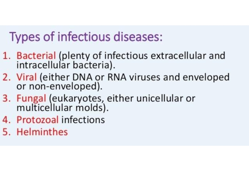
Infections are caused by infectious agents including
Bacteria
Viruses
Fungi (ringworm)
Protozoa
Macroparasites (nematodes, tapeworms, helminths)
ProtozoaMacroparasites (nematodes, tapeworms, helminths)")

Слайд 6PATHOGENICITY
Pathogenicity is a specific sign of the pathogen, its potential to
cause a specific infectious process under favorable conditions. On this basis, all states of microbes are subdivided into pathogenic, opportunistic and saprophytes. Pathogenicity and virulence are different concepts. A microorganism is considered virulent if it, when introduced into the animal's organism, even in small doses, causes the development of an infectious process.

Слайд 7VIRULENCE
Virulence is the degree of pathogenicity of a particular microorganism, i.e.
this is an individual characteristic. Virulence is a quantity that is measured (e.g., minimum lethal dose DLM, median lethal dose LD50).

Слайд 8SPECIFICITY
Each infectious disease causes a specific pathogen. So, the causative agent
of plague causes plague, cholera - cholera, etc. Infections (for example, purulent-inflammatory processes) caused by various microbes are known. On the other hand, one causative agent (for example, streptococcus) is capable of causing various lesions.

Слайд 9ORGANOTROPICITY
Organotropicity is the defeat of cells, tissues and organs that are
most suitable for their biochemical properties for life support of this type of microorganism.

Слайд 10VIRULENCE FACTORS
1. Adhesion to cells
Many bacteria for infection of certain cells
of the body, for example, intestinal epithelium should attach to them. It was found that a large number of host cell molecules, in particular, and the receptors of bacteria (proteins of the outer bacterial membrane) are involved in this process.

Слайд 11VIRULENCE FACTORS
2. Invasiveness
Some virulent bacteria produce proteins that destroy cell membranes
or stimulate phagocytosis of host cells. These virulence factors allow bacteria to enter the host's body through the layers of cells that come in contact with the pathogen, whether they are cells of the outer covers of plants or animals or layers of epithelium of internal organs.

Слайд 12VIRULENCE FACTORS
3. Colonization is the process of multiplication of microbes at
the site of adhesion. Colonization provides the accumulation of microorganisms to such a critical concentration that can cause a pathological effect.

Слайд 13VIRULENCE FACTORS
4. Suppression of the immune response
Many bacteria release virulence factors
that inhibit the body's immune system. For example, bacteria secrete proteins that attach to host antibodies. Another type of substance that inhibits the immune response is the polysaccharide capsule surrounding the cell. These polysaccharides complicate phagocytosis of bacteria by specialized cells of the immune system (macrophages) and lymphocytes.

Слайд 14VIRULENCE FACTORS
5. Toxins
Many virulence factors are proteins that the pathogen produces
and then secretes into the environment and which causes damage to the host tissues. For example, with food poisoning it is the toxins that cause the symptoms of the disease.


Слайд 18FORMS OF INFECTION PROCESSES
Infectious disease
Bacteriocarrier
Chronic (presence of pathogens for several
months or even years)
Transitory (short-term (most often - once) excretion of the pathogen in the absence of clinical manifestations of the disease)
Sharp (a consequence of a recent illness)
Transitory (short-term (most often - once) excretion of the pathogen in the absence of clinical manifestations of the disease)
Sharp (a consequence of a recent illness)
Transitory")

Слайд 20REASONS OF ORAL INFECTIONS
injuries
a lack of vitamins and trace elements
a general
decrease in immunity
allergic reactions
infections
the presence of tartar deposits
poor hygienic condition of the cavity
allergic reactions
infections
the presence of tartar deposits
poor hygienic condition of the cavity

Слайд 21BIOLOGICAL RESEARCH METHODS
Biological research methods are aimed at determining the presence
of pathogen toxins in the test material and on the detection of the causative agent. Methods include infecting laboratory animals with the test material, followed by isolation of a pure pathogen culture or establishing the presence of a microbial toxin and its nature. The method is highly sensitive, can be used in the early stages of the disease, but is not always available, expensive, long-lasting, unsafe.

Слайд 22OBJECTIVES OF BIOLOGICAL METHOD
1. Diagnosis of infectious diseases.
2. Identification of pure
culture.
3. Definition of virulence.
4. Isolation and identification of exotoxins.
5. Cultivation of viruses.
6. Reception of immunopreparations.
7. Checking the harmlessness and effectiveness of medications (including chemotherapy drugs, immunopreparations) and others.
3. Definition of virulence.
4. Isolation and identification of exotoxins.
5. Cultivation of viruses.
6. Reception of immunopreparations.
7. Checking the harmlessness and effectiveness of medications (including chemotherapy drugs, immunopreparations) and others.

Слайд 23STAGES OF BIOLOGICAL METHOD
1. Material intake.
2. Material processing.
3. The choice of
a laboratory animal.
4. Infection of animals by one of the methods.
5. Registration of signs of illness of the infected animal or its death.
6. Intravital sampling of material from the animal and carrying out bacteriological and serological studies, setting an allergic test.
7. Autopsy, study of pathoanatomical and pathomorphological pattern. Preparation of smears-prints from internal organs.
8. Identification of the selected culture.
9. Conclusion on the results of the study.

Слайд 24TASK 1
STAINING BY BURRY-HINS METHOD
The Burry-Hins method is used to color
capsular bacteria and is based on the fact that the capsule does not perceive the dyes. The capsule is detected by negative contrasting backgrounds over the Burry. For this, black mascara is mixed into culture and dried. After that, for fixation in the flame of the burner, stain the bodies of microbial cells according to Hins – aqueous fuchsine for 1 minute and rinse with water for 5-10 seconds.
NB! As a result, a colorless capsule and red microbial bodies are clearly visible on a dark background.
NB! As a result, a colorless capsule and red microbial bodies are clearly visible on a dark background.

Слайд 25TASK 1
STAINING BY BURRY-HINS METHOD
Klebsiella pneumonia.
A smear of pure culture.
Burri-Hins staining

Слайд 26TASK 2
PETRI DISH WITH S. AUREUS INOCULATED ON BLOOD AGAR
Hemolysis or
haemolysis is the rupturing (lysis) of the red blood cells (erythrocytes) and the release of their contents (cytoplasm) into the surrounding fluid (e.g. blood plasma). Hemolysis may occur in vivo or in vitro (inside or outside the body).
Hemolysins damage the host cytoplasmic membrane, causing cell lysis and death. The activity of these toxins is most easily observed with assays involving the lysis of the red blood cells (erythrocytes).
Hemolysins damage the host cytoplasmic membrane, causing cell lysis and death. The activity of these toxins is most easily observed with assays involving the lysis of the red blood cells (erythrocytes).

Слайд 27TASK 2
PETRI DISH WITH S. AUREUS INOCULATED ON BLOOD AGAR
The pathogenic
properties of staphylococci are due to the ability to produce exotoxins and enzymes of aggression. They cause lysis of erythrocytes, have a lethal and necrotic effect. The most studied hemolytic properties of toxins, so they are often called hemolysins. The hemolytic ability of staphylococci can be determined by inoculation on blood agar, in which 18-24 hours around the staphylococcus colony, the hemolysis zone is visible.

Слайд 28TASK 2
PETRI DISH WITH S. AUREUS INOCULATED ON BLOOD AGAR
1 –
Staphylococcus colonies
2 – Hemolysis zones
3 – Blood agar
2 – Hemolysis zones
3 – Blood agar
1
2
3

Слайд 29TASK 3 TEST TUBES WITH NORMAL RABIT PLASMA AND WITH S. AUREUS
PLAQUE
Plasma-coagulant activity of the culture is checked by inoculation in tubes with 0.5 ml of citrated rabbit plasma at a dilution of 1: 4. Pathogenic staphylococci coagulate the plasma for 2-24 h under the conditions of a thermostat. Accounting is performed after 24 hours by the formation of a small jelly-like clot on the bottom of the tube.

Слайд 30TASK 3 TEST TUBES WITH NORMAL RABIT PLASMA AND WITH S. AUREUS
PLAQUE
Plasmacoagulase positive
Plasmacoagulase negative

Слайд 31TASK 4
S- AND R-FORMS OF COLONIES
S-form
Round
Smooth
Even edges
Shiny surface
R-form
Irregular in shape
Rough
Serrated edges