- Главная
- Разное
- Дизайн
- Бизнес и предпринимательство
- Аналитика
- Образование
- Развлечения
- Красота и здоровье
- Финансы
- Государство
- Путешествия
- Спорт
- Недвижимость
- Армия
- Графика
- Культурология
- Еда и кулинария
- Лингвистика
- Английский язык
- Астрономия
- Алгебра
- Биология
- География
- Детские презентации
- Информатика
- История
- Литература
- Маркетинг
- Математика
- Медицина
- Менеджмент
- Музыка
- МХК
- Немецкий язык
- ОБЖ
- Обществознание
- Окружающий мир
- Педагогика
- Русский язык
- Технология
- Физика
- Философия
- Химия
- Шаблоны, картинки для презентаций
- Экология
- Экономика
- Юриспруденция
Transcriptional regualtion. Repression: Hypoxic Genes in Yeast презентация
Содержание
- 1. Transcriptional regualtion. Repression: Hypoxic Genes in Yeast
- 2. Regulation of gene expression Almost as important
- 3. A yeast model for repression of gene
- 4. Isolation of mutations affecting ANB1 repression Inversion
- 5. Characterizing mutations in ANB1 regulation cis-acting mutations
- 6. Characterization of the rox1 mutation The initial
- 7. Cloning of the rox1 mutation De-repression
- 8. Cloning of rox1 mutation (2) rox1 mutant
- 9. Cloning of rox1 mutation (3) Grow rox1,
- 10. The Rox1 protein is the repressor of
- 11. Rox1p requires Ssn6/Tup1 for repression In a
- 12. Model of protein and nucleosome interactions at
- 13. Ssn6/Tup1 recruit HDACs to establish a repressive
- 14. URS HYPOXIC Genes
- 15. 2. Ssn6/Tup1 interacts with the RNA poymerase
- 16. Oxygen regulation in yeast
- 18. Promoter analysis What determines the efficiency
- 20. 3 .5
- 21. Role of position for repressor efficiency
- 22. through OpA
- 23. The sequence TGCCT is responsible for stronger repression from OpA
- 24. Insertion of the conserved sequence adjacent to
- 25. MOT3 (Modulator Of Transcription): Mutant derepresses
- 26. Electrophoretic mobility shift assay (EMSA) Used in
- 27. EMSA - Principle DNA with binding site DNA – protein complex (High molecular weight, bulky)
- 28. Rox1
- 29. A mot3 deletion causes mild derepression of
- 30. How does Mot3p exert its effect on
- 31. +R1 -R1 20ng Mot3
- 32. A micrococcal nuclease (MNase ) digest
- 33. Mot3 affects the chromatin structure of the
- 34. MCNase generated digestion pattern is dependent on histone N-termini MNAse
- 35. Summary Operator efficiency: -
- 36. A Model Fungal Gene Regulatory Mechanism: The
- 37. GAL mutant phenotypes: GAL1, GAL7, GAL10, MEL1,
- 38. The GAL structural genes GAL1, GAL7, GAL10,
- 39. GAL4 and GAL80 are regulatory proteins gal4-
- 40. promoter Gal4p
- 41. promoter 2. Gal4p is the activator of
- 42. Galactose promoter
- 43. promoter Galactose
- 44. How can we distinguish between the two
- 45. Scenario 2 is correct: the gal4-/gal80- mutant
- 46. Cloning of the genes gal4- uninducible, cannot
- 47. The Gal4p Activator The Gal4 protein is
- 48. Gal4p binds UAS sequences in the regulatory
- 49. lacZ UAS Deletion analysis of
- 50. Gal4p is a modular protein H2N DNA bd Act Activ./Gal80 ia bd
- 51. lacZ UAS VP16
- 52. Expression of GAL4 itself is regulated by
- 53. The galactose sensor: Gal3p Gal3p is a
- 54. In a nutshell…. Glucose (repressed):
- 55. other carbon source than Glucose (derepressed): GAL genes UAS
- 56. Galactose: UAS +Galactose GAL genes
- 57. What is the mechanism of transcriptional activation
- 58. UAS GAL genes TATA
- 59. Micrococcal nuclease digest of chromatin
- 60. Nucleosome Perturbation via recruitment of Histone Acetyl-transferases
- 61. B. Gal4p interacts directly with the TATA-
- 62. Relevance of the Gal regulation research today?
- 63. Galactose induction can be utilized to overexpress
- 64. Three expression levels: Repressed (2% glucose) ?
- 65. Similar: Oleate induction: Oleate induced genes are
- 66. Expression from inducible promoters allows investigation of
Слайд 1Repression:
Hypoxic Genes in Yeast
Rox1p, Tup1p, Ssn6/Cyc8p and Mot3p
Transcriptional regulation

Слайд 2Regulation of gene expression
Almost as important as the genetic repertoire itself
The
Five (six?)regulatory levels:
(DNA copy number)
Transcription
mRNA stability
Translation
Post-translational modifications
Protein stability

Слайд 3A yeast model for repression of gene transcription
The transcription of the
ANB1 codes for the essential eIF-5A protein involved in translation initiation or mRNA export from the nucleus
In the presence of oxygen, ANB1 is strongly repressed, and an aerobic counterpart, TIF51A, which codes for and almost identical protein, is activated. Yeast needs the eIF-5A protein from one or the other gene to survive
ANB1 is closely linked to the yeast oxygen-activatedCYC1 gene, which codes for the Iso-1-cytochrome that is required for respiration

Слайд 4Isolation of mutations affecting ANB1 repression
Inversion of regulatory region
Part of the
This manipulation puts CYC1 under the control of the ANB1 regulatory region
CYC1 is highly repressed; a strain that carries a cyc1 mutation on its genomic DNA and the plasmid with the inversion cannot grow on non-fermentable carbon sources, because no functional iso-1-cytochrome c is made
This strain was treated with UV light or EMS (ethylmethane sulfonate, and alkylating agent) to obtain mutants in which repression by oxygen is relieved

Слайд 5Characterizing mutations in ANB1 regulation
cis-acting mutations (mutations on the plasmid in
mating the mutant strain to the parental strain (cyc1 Δ); cis-acting mutations should act dominant (? diploid should remain respiratory competent), trans-acting loss-of-function mutants should be recessive (diploid should be unable to respire)
Growing cells on non-selective media (to lose the plasmid; 5-10% loss per generation) and re-transforming the mutant with the original plasmid (mutants in trans-acting protein factors should still be mutant? able to respire)
- Mutants were sorted into complementation groups
 were")
Слайд 6Characterization of the rox1 mutation
The initial rox1 mutant displayed de-repression of
Genetic analysis indicated the mutation was in one gene

Слайд 7
Cloning of the rox1 mutation
De-repression of hypoxic genes does not have
Creation of a reporter construct, integration into the URA3 locus of the rox1 mutant strain
The resulting strain is ura3- and expresses the lacZ gene product (β-galactosidase) constitutively
ANB1/lacZ
ANB1 promoter
URA3
UR
A3
ANB1/lacZ
ANB1 promoter
UR
A3
Restriction fragment from plasmid
(select for FOA resistance)

Слайд 8Cloning of rox1 mutation (2)
rox1 mutant cells with integrated ANB1-lacZ fusion
rox1 mutant cells with integrated ANB1-lacZ fusion on medium containing X-gal")
Слайд 9Cloning of rox1 mutation (3)
Grow rox1, ura3::ANB1-lacZ mutant cells
Plate on SC-
Screen for white colonies
lacZ expression from ANB1-lacZ fusion repressed by ROX1 gene from library plasmid ? no β-galcatosidase activity
Grow rox1, ura3::ANB1-lacZ mutant cellsPlate on SC- Ura, X-galScreen for white")
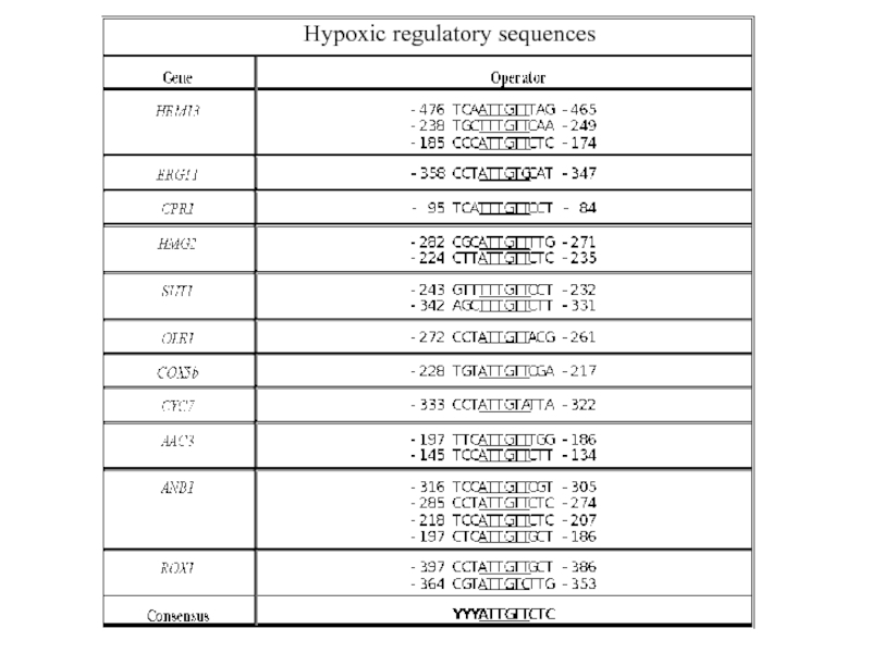
Слайд 10The Rox1 protein is the repressor of hypoxic genes
Rox1p is a
The DNA – binding domain has high similarity to the DNA-binding domain of the human Sry gene involved in sex-determination and to proteins conferring resistance to the drug cis-platin used in cancer therapy
The DNA – binding domain is roughly L-shaped and introduces 90o bends into DNA
Rox1 binding site consensus:
YYYATTGTTCTC

Слайд 11Rox1p requires Ssn6/Tup1 for repression
In a similar screen, mutations in the
Sequence analysis revealed that ROX4=TUP1 and ROX5=SSN6/CYC8
Rox1p is dependent on Ssn6/Tup1 for its repression activity and recruits the Ssn6/Tup1 complex to the target promoters of the hypoxic genes
The Tup1/Ssn6 repression complex consists of one Ssn6p subunit and three or four Tup1 subunits

Слайд 12Model of protein and nucleosome interactions at the RNR3 promoter. A,
Tup1/Ssn6 interacts with nucleosomes to form a repressive chromatine structure
B. Li and J. C. Reese
Ssn6-Tup1 Regulates RNR3 by Positioning Nucleosomes and Affecting the Chromatin Structure at the Upstream Repression Sequence
J. Biol. Chem, September 7, 2001; 276(36): 33788 - 33797.

Слайд 13Ssn6/Tup1 recruit HDACs to establish a repressive chromatin structure
Tup1 has been
Histone deacetylation causes tighter association of Histones with DNA due to the positive charge of K (Lysine) and R (Arginine) residues in the N-terminal tails of Histones H3 and H4
Tup1 has also been demonstrated to directly interact with hypo- (under-) acetylated H3 and H4


Слайд 152. Ssn6/Tup1 interacts with the RNA poymerase II mediator complex
Figure 3.
A. J. Courey and S. Jia
Transcriptional repression: the long and the short of it
Genes & Dev., November 1, 2001; 15(21): 2786 - 2796.


Слайд 18Promoter analysis
What determines the efficiency of repression?
- Sequence of repressor
- Number of operators/ repressor binding sites
- Position?
- Modulating factors?

Слайд 203
.5
OpA in OpB site
0
.86
43
50
ANB1/lacZ
OpA
OpB
TATA
31 bp
)
)
)
$
)
)
Organization of the Operators in the ANB1 Regulatory Region
Rox1 Binding Site Rearrangements
))$))Organization of")


Слайд 24Insertion of the conserved sequence adjacent to the OpA 5’ Rox1

Слайд 25MOT3 (Modulator Of Transcription):
Mutant derepresses DAN1 (Delayed Anaerobic) and ANB1
Sertil O,
- poorly characterized transcriptional regulator
- deletion with weak mutant phenotype (marginally slower growth; positive and negative effects on transcription)
- DNA binding protein with two C2H2 zinc fingers, localized to the nucleus
- binding site is T(A>G)CCT(G>T>A)
site in OpA: TGCCT
Does the Mot3 protein bind OpA?
:Mutant derepresses DAN1 (Delayed Anaerobic) and ANB1Sertil O, Kapoor R, Cohen BD,")
Слайд 26Electrophoretic mobility shift assay (EMSA)
Used in analysis of DNA binding properties
Binding target (DNA or RNA, often a short oligomer containing protein binding sites) is labelled radioactively
Binding of protein to DNA results in retardation of the migration of the labelled DNA band
Used in analysis of DNA binding properties of proteinsBinding target (DNA")
")
Слайд 28Rox1
Mot3
The Mot3 protein
promoter
- 1 5 1 1 1 5 5 5
- - - 5x 20x - 5x 20x - - 20x -
- - - - - 20x - - 20x - - 20x
competitor DNA
labelled DNA
OpA
OpA (-Mot3 site)
OpA
Mot3 site
Rox1 site
Does Mot3p play a role in ANB1 repression in vivo?

Слайд 29A mot3 deletion causes mild derepression of ANB1
Northern blot probing for

Слайд 30How does Mot3p exert its effect on repression?
1. Interaction with Rox1p?
2. Interaction with the Ssn6/Tup1 general repression complex?
- establishment complex formation?
- aiding repression function?
2. Interaction with")
Слайд 31
+R1
-R1
20ng
Mot3
25ng Rox1
MBP
Free
DNA
20ngGST-
Mot3
Mot3 and Rox1 do not bind
+R1
Rox1 site
Mot3 site
-R1
labelled DNA
competitor DNA

Слайд 32A micrococcal nuclease (MNase ) digest reveals chromatin structure of
Operator
ANB1
TATA
Operator
ANB1
Radioactive probe anneals to 3’ end of DNA
DNA fragments of different lenghts created by MNase digest of unprotected DNA
Protectionm from MNase cleavage results in the disappearance of certain DNA fragments
 digest reveals chromatin structure of regulatory regionsOperatorANB1TATAOperatorANB1Radioactive probe anneals to")
Слайд 33Mot3 affects the chromatin structure of the ANB1 promoter in a
manner as Tup1, Ssn6 and Rox1
MNAse


Слайд 35Summary
Operator efficiency:
- operator orientation and position relative to the TATA
- the sequence TGCCT between OpA binding sites is responsible
for higher repression efficiency of OpA compared to OpB
- the TGCCT sequence improves repression from OpB when inserted
- the TGCCT sequence is bound specifically by the transcription
factor Mot3, a zinc finger protein protein that has been reported affect the expression of various other genes
- deletion of the MOT3 gene causes partial derepression of hypoxic genes

Слайд 36A Model Fungal Gene Regulatory Mechanism: The GAL genes of Saccharomyces
GAL genes: involved in Galactose metabolism
Early results:
GAL genes are repressed in the presence of glucose
“ “ “ derepressed in presence of other carbon sources
most GAL genes induced about 1000 fold upon addition of galactose to media (as long as no glucose available)

Слайд 37GAL mutant phenotypes:
GAL1, GAL7, GAL10, MEL1, (GAL5): If mutant, cells cannot
GAL2: Mutant cells cannot utilize galactose, but all enzymatic activities are present in cell extract
GAL4: Mutant cells cannot utilize galactose, none of the enzymatic activities are present in cell extract
GAL3: In combination with mutation in any one mutation in GAL1, GAL7, GAL10, MEL1 (GAL5), cells cannot utilize galactose, and all of the enzymatic activities are missing
GAL80: All enzymatic activities are constitutively expressed
: If mutant, cells cannot utilize galactose; a specific")
Слайд 38The GAL structural genes
GAL1, GAL7, GAL10, induced >1000x on galactose
MEL1 induced
(GAL5 ~ 3-4 x)

Слайд 39GAL4 and GAL80 are regulatory proteins
gal4- : uninducible (recessive)
gal80- : constitutive
Two (very simplified!!!) models for mode of action:
1. Gal80p is a repressor of the GAL genes; Gal4p inactivates Gal80p in the presence of galactose
promoter
Gal80p
Gal4p
gal80- : constitutive (recessive)Two (very simplified!!!) models")

Слайд 41promoter
2. Gal4p is the activator of the GAL genes; Gal80p is


Слайд 44How can we distinguish between the two models?
Epistasis analysis of pathway
What
promoter
Galactose
promoter
Galactose
Gal4p
1.
2.

Слайд 45Scenario 2 is correct: the gal4-/gal80- mutant is uninducible
Gal4p is an
Recessiveness characteristic for loss of function mutant
GAL4c mutation: constitutive (cannot interact with GAL80) ? dominant mutation
GAL80u mutation: uninducible; (does not respond to galactose)? dominant

Слайд 46Cloning of the genes
gal4- uninducible, cannot grow on plates with galactose
gal80- constitutive: use of inhibitor 2-deoxygalactose (kills cells that are able to metabolize galactose) ? transform cells on media with inhibitor (+ other carbon source) and select for survivors

Слайд 47The Gal4p Activator
The Gal4 protein is a DNA - binding transcriptional
H2N
COOH
DNA bd
Act
Zn2+ finger domain
+ dimerization domain
Gal80p binding domain
+ transcriptional activation domain

Слайд 48Gal4p binds UAS sequences in the regulatory region of GAL structural
GAL genes
UAS
UAS: upstream activation sequence
TATA – box: AT-rich sequence required for transcription machinery assembly

Слайд 49
lacZ
UAS
Deletion analysis of promoter region identified Gal4p binding sites
Gal4p binding site:
5’
3’TCAGG/CAGGC-5’
Site is promiscuous (can function if front of many genes
Orientation & position independent (symmetrical site, wide range of upstream region from where it can exert transactivation)
“Gal4 17-mer”


Слайд 51
lacZ
UAS
VP16
Activation domain (758-881)can activate independently of the rest of the protein
lacZ
lexA binding site
Gal4 Activ.
Gal4 bd
lexA bd
DNA binding domain (1-174) can bind DNA without the rest of the protein and can target a heterologous activation domain (VP16, viral activation domain) to promoters with a GAL UAS and exert transcriptional activation
can activate independently of the rest of the protein if fused to a")
Слайд 52Expression of GAL4 itself is regulated by glucose
Under high glucose concentrations,

Слайд 53The galactose sensor: Gal3p
Gal3p is a protein with high similarity (homology)
No enzymatic activity
In the presence of galactose, Gal3p binds the sugar and removes the Gal80p repressor from the Gal4p activator
 to galactokinaseNo enzymatic activityIn")
:Mig1pSsn6/Tup1GAL genesMig1pSsn6/Tup1UAS")
:GAL genesUAS")

Слайд 57What is the mechanism of transcriptional activation by Gal4p?
A. Gal4p activates
UAS
GAL genes
TATA
RNApolII
Mediator complex
TBP


Слайд 59Micrococcal nuclease digest of chromatin
UAS
GAL genes
TATA
UAS
GAL genes
Radioactive probe anneals to 3’
EcoRV
EcoRV

Слайд 60Nucleosome Perturbation via recruitment of Histone Acetyl-transferases (HATs)?
Histones have positively charged
Gal4 is suspected to recruit HATs (e.g Gcn5p/SAGA complex) to the promoters of the GAL genes and thereby locally disrupt histone-DNA interaction
?Histones have positively charged N-terminal tails (K/R –")
Слайд 61B. Gal4p interacts directly with the TATA- binding protein or the
polymerase II complex
UAS
GAL genes

Слайд 62Relevance of the Gal regulation research today?
General understanding of basic molecular
Model for the functioning of biological regulatory circuits
A general mechanism for network-dosage compensation in gene circuits. Acar M, Pando BF, Arnold FH, Elowitz MB, van Oudenaarden A. Science. 2010 Sep 24;329(5999):1656-60

Слайд 63Galactose induction can be utilized to overexpress heterologous genes
Genes of interest
YFG1
GAL1 promoter (4 Gal4p binding sites)

Слайд 64Three expression levels:
Repressed (2% glucose) ? no expression
Derepressed (2% Raffinose, 3%
Activated (2% Galactose) ? high expression
Useful for:
Overexpression for purification
Multicopy effect studies
Study of essential genes (genes for which deletions are lethal)
 ? no expressionDerepressed (2% Raffinose, 3% Glycerol) ? intermediate expressionActivated")
Слайд 65Similar: Oleate induction:
Oleate induced genes are involved in peroxisomal proliferation and
Activator is a heterodimer of the Oaf1p/Pip2p activators which bind to oleate response elements (OREs)
The ORE consensus is currently viewed as two inverted CGG triplets spaced by 14 (formerly 15) to 18 intervening nucleotides (N), i.e. CGGN3TNAN8-12CCG
Currently, the plasmid available has the promoter and terminator sequences of the oleate-induced CTA1 (peroxisomal catalase) gene
CTA1 is glucose repressed similar to the GAL genes
Three expression levels:
Repressed (2% Glucose)
Derepressed (2% Raffinose, 3% Glycerol)
Activated (0.2% oleate, 0.02% Tween, 0.05% Glucose)

Слайд 66Expression from inducible promoters allows investigation of essential genes
Essential genes are
Deletions of these genes are inviable, deletion are only viable as heterozygous diploids, or deletion strains have to carry a plasmid with a wild type copy of the gene
Shuffling in plasmids carrying mutant partial function alleles is one way of investigating the function
Introduction of plasmids with the essential gene expressed from an inducible promoter allow more precise investigation