- Главная
- Разное
- Дизайн
- Бизнес и предпринимательство
- Аналитика
- Образование
- Развлечения
- Красота и здоровье
- Финансы
- Государство
- Путешествия
- Спорт
- Недвижимость
- Армия
- Графика
- Культурология
- Еда и кулинария
- Лингвистика
- Английский язык
- Астрономия
- Алгебра
- Биология
- География
- Детские презентации
- Информатика
- История
- Литература
- Маркетинг
- Математика
- Медицина
- Менеджмент
- Музыка
- МХК
- Немецкий язык
- ОБЖ
- Обществознание
- Окружающий мир
- Педагогика
- Русский язык
- Технология
- Физика
- Философия
- Химия
- Шаблоны, картинки для презентаций
- Экология
- Экономика
- Юриспруденция
The Cytoskeleton: Intermediate Filaments and Microtubules презентация
Содержание
- 1. The Cytoskeleton: Intermediate Filaments and Microtubules
- 2. The Cytoskeleton Includes Dynamic Networks Of Microfilaments And Microfilaments
- 3. The cytoskeleton consists of three major types
- 4. YPET – MAP R – MT PLUS
- 5. Intermediate Filaments are non- dynamic and
- 6. Intermediate Filaments polymerize to form strong rope-like
- 7. The inner side of the nuclear envelope
- 8. Intermediate filament networks flare out from the
- 9. Microtubules Make Up Dynamic Networks
- 10. Microtubules serve four functions: To give
- 11. Microtubules Are Made Of Tubulin Protofilaments
- 12. Microtubules as seen by Electron Microscopy
- 13. Microtubules are stabilized by capping at their
- 14. The centrosome consists of centrioles surrounded by
- 15. Microtubule assembly at plus end is governed
- 16. Catastrophic Disassembly can occur if growth
- 17. DYNAMIC INSTABILITY IN A MICROTUBULE ASTER
- 18. MICROTUBULE DYNAMICS SEEN WITH FLUORESCENT PLUS END PROTEINS
- 19. MICROTUBULE DYNAMICS SEEN WITH FLUORESCENT PLUS END PROTEINS
- 20. Microtubule associated proteins also stabilize microtubules. Acetylation and tyrosylation do too.
- 21. Drugs can stabilize or destabilize microtubules;
- 22. Motor proteins “walk” on microtubules and microfilaments
- 23. Kinesin, like myosin, hydrolyzes ATP as it
- 24. MOTOR PROTEINS MOVE VESICLES ON MICROTUBULE TRACKS
- 25. MOTOR PROTEINS MOVE VESICLES ON MICROTUBULE TRACKS
- 26. Direction of vesicle Transport on microtubules FIBROBLAST NEURON Movement of pigment granules on MTs
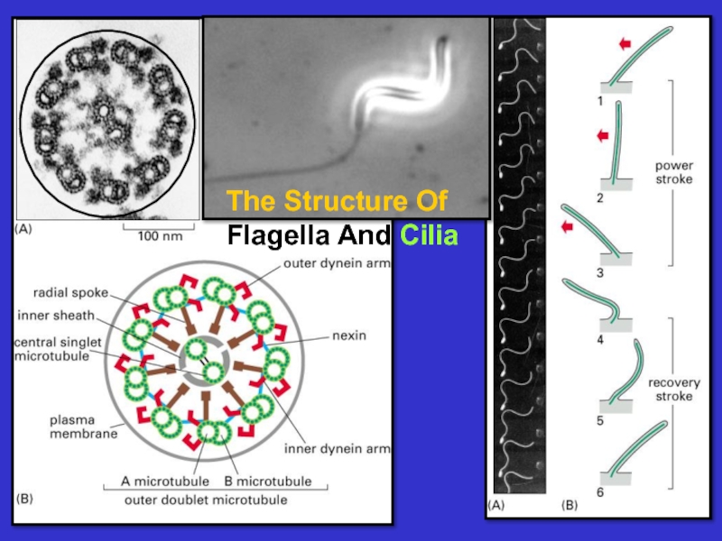
- 27. Cilia And Flagella: A Different Form Of Motility
- 29. Dynein provides Motive force to move one
- 30. Dynein Motors cause microtubule sliding in
Слайд 1Lecture 20:
The Cytoskeleton:
Intermediate Filaments and Microtubules
Essential
Cell Biology
Fourth Edition
Chapter 17


Слайд 3The cytoskeleton consists of three major types
of filaments plus many
filament-associated
proteins including molecular motors
proteins including molecular motors
Microfilaments – composed of actin, these
filaments form dynamic networks that form
the basis for cell shape and movement
Microtubules – composed of tubulin, these
tubules act as tracks on which to move
vesicles and organelles. They also form the
basis of cilia and flagella. They are dynamic.
Intermediate filaments – composed of proteins
that associate to form rope-like structures
that are of high mechanical strength. They
position organelles and form a strong, long
lasting cell superstructure.

Слайд 4YPET – MAP R – MT PLUS END
KERITAN – INTERMEDIATE FIL.
ACTIN
– STRESS FIBERS
ACTININ – STRESS FIBERS
VIMENTIN – INTERMEDIATE
TUBULIN - MICROTUBULES
Cytoskeletal
Networks
Containing
Fluorescent
Proteins
Fluorescence
Microscopy allows
Visualization
Of cytoskeletal
Networks

Слайд 5Intermediate
Filaments
are non-
dynamic and
structural.
They position
the nucleus
and insert into
Desmosomes
to hold
neighboring
cells
together.

Слайд 6Intermediate
Filaments
polymerize
to form strong
rope-like fibers. The basic
structural unit is a coiled-coil
dimer.
These
fibers are symmetric
fibers are symmetric

Слайд 7The inner side of the nuclear envelope
is lined by a network
of intermediate
filaments called lamins. They serve as
an anchoring site for chromosomes as
well as for intermediate filament networks
that extend from the nucleus out into the
cytoplasm.
filaments called lamins. They serve as
an anchoring site for chromosomes as
well as for intermediate filament networks
that extend from the nucleus out into the
cytoplasm.

Слайд 8Intermediate filament networks flare out from the nucleus and insert into
plasma membrane junctions called desmosomes. Desmosomes connect the intermediate filaments networks of neighboring cells forming a strong mechanical bond that keeps the cells from being pulled apart.


Слайд 10Microtubules serve four functions:
To give shape to the cell.
Example: nerve axons
contain numerous micro-
tubules along their length. If disrupted the axon
shrivels.
2. To provide “tracks” on which to move
vesicles carrying cargo.
Example: pigment granules move outward
and inward from cell center using microtubules.
3. To form the mitotic spindle which separates
chromosomes during mitosis and meiosis.
To form flagella and cilia – whip like
structures that propel cells.


Слайд 12Microtubules as seen by Electron
Microscopy
1) thin section
2) freeze dried
And platinum
Shadowed
 thin section2) freeze driedAnd platinumShadowed")
Слайд 13Microtubules are stabilized by capping at their
Plus and minus ends. Centrosomes
and
Microtubule organizing centers (MTOCs) cap the
minus end; special membrane-associated proteins
cap the plus end.
Microtubule organizing centers (MTOCs) cap the
minus end; special membrane-associated proteins
cap the plus end.

Слайд 14The centrosome consists of centrioles surrounded by a “protein cloud”. Minus
ends of microtubules are capped by gamma tubulin rings and the centrosome serves as a microtubule organizing center (MTOC).

Слайд 15Microtubule assembly
at plus end is governed
by GTP hydrolysis; GTP-
tubulin is
required for
polymerization;
But after hydrolysis,
GDP-tubulin
favors depolymerization
polymerization;
But after hydrolysis,
GDP-tubulin
favors depolymerization

Слайд 16Catastrophic
Disassembly can occur if growth at the plus end stops
or is slow; but
the microtubule starts to grow at this end again.
the microtubule starts to grow at this end again.


Слайд 20Microtubule associated proteins also stabilize microtubules.
Acetylation and
tyrosylation do too.

Слайд 21Drugs can stabilize or destabilize microtubules;
Taxol stabilizes existing mts; cholchicine
destabilizes microtubules by monomer binding


Слайд 23Kinesin, like myosin, hydrolyzes ATP as it walks
During this process chemical
energy is
transformed into mechanical energy, hence the name motor protein.
transformed into mechanical energy, hence the name motor protein.

Слайд 24MOTOR PROTEINS MOVE VESICLES ON MICROTUBULE TRACKS –
A CONFORMATIONAL CYCLE
THAT HYDROLYZES ATP


Слайд 26Direction of vesicle
Transport on microtubules
FIBROBLAST
NEURON
Movement of pigment granules on MTs