- Главная
- Разное
- Дизайн
- Бизнес и предпринимательство
- Аналитика
- Образование
- Развлечения
- Красота и здоровье
- Финансы
- Государство
- Путешествия
- Спорт
- Недвижимость
- Армия
- Графика
- Культурология
- Еда и кулинария
- Лингвистика
- Английский язык
- Астрономия
- Алгебра
- Биология
- География
- Детские презентации
- Информатика
- История
- Литература
- Маркетинг
- Математика
- Медицина
- Менеджмент
- Музыка
- МХК
- Немецкий язык
- ОБЖ
- Обществознание
- Окружающий мир
- Педагогика
- Русский язык
- Технология
- Физика
- Философия
- Химия
- Шаблоны, картинки для презентаций
- Экология
- Экономика
- Юриспруденция
The biological perspective презентация
Содержание
- 1. The biological perspective
- 2. Learning Objectives 2.1 What are the nervous system,
- 3. Overview of Nervous System Nervous system an
- 4. Structure of the Neuron Neuron the basic
- 5. Structure of the Neuron Parts of a
- 6. Figure 2.2 The Structure of the Neuron The electronmicrograph on the left shows myelinated axons.
- 7. Other Types of Brain Cells Glial cells
- 8. Other Types of Brain Cells Myelin: fatty
- 9. Generating the Message: Neural Impulse Ions: charged
- 10. Generating the Message: Neural Impulse All-or-none: a
- 11. Figure 2.3 The Neural Impulse Action Potential
- 12. Figure 2.3 (continued) The Neural Impulse Action
- 13. Communication Between Neurons Sending the message to
- 14. Neuron Communication Synaptic vesicles: sack-like structures found
- 15. Neuron Communication synapse/synaptic gap: microscopic fluid-filled space
- 16. Figure 2.4 Reuptake of Dopamine
- 17. Neuron Communication Neurons must be turned ON
- 18. Neuron Communication Chemical substances can affect neuronal
- 20. Cleaning up the Synapse reuptake: process by
- 21. Figure 2.5 An Overview of the Nervous System
- 22. Central Nervous System Central nervous system (CNS):
- 23. The Reflex Arc: Three Types of Neurons
- 24. The Reflex Arc: Three Types of Neurons
- 25. The Reflex Arc: Three Types of Neurons
- 26. Peripheral Nervous System Peripheral nervous system (PNS):
- 27. Figure 2.7 The Peripheral Nervous System
- 28. Somatic Nervous System Soma = “body” Somatic
- 29. Somatic Nervous System Somatic nervous system (cont’d)
- 30. Autonomic Nervous System Autonomic nervous system (ANS)
- 31. Autonomic Nervous System Autonomic Nervous System (ANS)
- 32. Figure 2.8 Functions of the Parasympathetic and Sympathetic Divisions of the Nervous System
- 33. The Endocrine Glands Endocrine glands: glands that
- 34. Figure 2.9 The Endocrine Glands
- 35. The Endocrine Glands pituitary gland: gland located
- 36. The Endocrine Glands gonads: the sex glands;
- 37. Looking inside the Living Brain Clinical Studies
- 38. Looking inside the Living Brain Clinical Studies
- 39. Mapping Structure computed tomography (CT): brain-imaging method
- 40. Mapping Structure Mapping Function electroencephalogram (EEG): records
- 41. Mapping Structure Mapping Function (cont’d) single photon
- 42. Figure 2.12 Major Structures of the Human Brain
- 43. The Hindbrain The Hindbrain medulla: first large
- 44. The Hindbrain reticular formation (RF): area of
- 45. Figure 2.13 The Limbic System
- 46. Structures under the Cortex Limbic system: a
- 47. Structures under the Cortex Limbic System (cont’d)
- 48. Structures under the Cortex Limbic System (cont’d)
- 49. Cortex cortex: outermost covering of the brain
- 50. Cerebral Hemispheres cerebral hemispheres: the two sections
- 51. Figure 2.14 The Lobes of the Brain
- 52. Four Lobes of the Brain occipital lobe:
- 53. Four Lobes of the Brain parietal lobes
- 54. Figure 2.15 The Motor and Somatosensory Cortex
- 55. Four Lobes of the Brain temporal lobes:
- 56. Four Lobes of the Brain frontal lobes:
- 57. Association Areas of Cortex association areas: areas
- 58. Association Areas of Cortex Broca’s aphasia: condition
- 59. Association Areas of Cortex Wernicke’s aphasia: condition
- 60. Association Areas of Cortex spatial neglect: condition
- 61. Split-Brain Research Cerebrum: the upper part of
- 62. Split-Brain Research Split-Brain Research study of patients
- 64. Results of Split-Brain Research left side of
- 65. Attention-Deficit/Hyperactivity Disorder Causes of ADHD have highlighted

Слайд 2Learning Objectives
2.1 What are the nervous system, neurons, and nerves, and how
2.2 How do neurons use neurotransmitters to communicate with each other and with the body?
2.3 How do the brain and spinal cord interact, and what are some misconceptions about the brain, and what is neuroplasticity?
2.4 How do the somatic and autonomic nervous systems allow people and animals to interact with their surroundings and control the body’s automatic functions?
2.5 How do the hormones released by glands interact with the nervous system and affect behavior?
2.6 How do psychologists study the brain and how it works?
2.7 What are the different structures of the hindbrain and what do they do?
2.8 What are the structures of the brain that control emotion, learning, memory, and motivation?
2.9 What parts of the cortex control the different senses and the movement of the body?
2.10 What parts of the cortex are responsible for higher forms of thought, such as language?
2.11 How does the left side of the brain differ from the right side?
2.12 What are some potential causes of attention-deficit/hyperactivity disorder?

Слайд 3Overview of Nervous System
Nervous system
an extensive network of specialized cells that
Neuroscience
deals with the structure and function of neurons, nerves, and nervous tissue
relationship to behavior and learning
LO 2.1 What Are the Nervous System, Neurons, and Nerves?

Слайд 4Structure of the Neuron
Neuron
the basic cell that makes up the nervous
LO 2.1 What Are the Nervous System, Neurons, and Nerves?

Слайд 5Structure of the Neuron
Parts of a neuron
dendrites: branch-like structures that receive
soma: the cell body of the neuron, responsible for maintaining the life of the cell
axon: long, tube-like structure that carries the neural message to other cells
LO 2.1 What Are the Nervous System, Neurons, and Nerves?

Слайд 6Figure 2.2 The Structure of the Neuron The electronmicrograph on the left

Слайд 7Other Types of Brain Cells
Glial cells are grey fatty cells that:
provide support for the neurons to grow on and around
deliver nutrients to neurons
produce myelin to coat axons
LO 2.1 What Are the Nervous System, Neurons, and Nerves?

Слайд 8Other Types of Brain Cells
Myelin: fatty substances produced by certain glial
clean up waste products and dead neurons
LO 2.1 What Are the Nervous System, Neurons, and Nerves?

Слайд 9Generating the Message: Neural Impulse
Ions: charged particles
inside neuron: negatively charged
outside neuron:
Resting potential: the state of the neuron when not firing a neural impulse
Action potential: the release of the neural impulse consisting of a reversal of the electrical charge within the axon
allows positive sodium ions to enter the cell
LO 2.1 What Are the Nervous System, Neurons, and Nerves?

Слайд 10Generating the Message: Neural Impulse
All-or-none: a neuron either fires completely or
Return to resting potential
LO 2.1 What Are the Nervous System, Neurons, and Nerves?

Слайд 11Figure 2.3 The Neural Impulse Action Potential In the graph below, voltage

Слайд 12Figure 2.3 (continued) The Neural Impulse Action Potential In the graph below,
 The Neural Impulse Action Potential In the graph below, voltage readings are")
Слайд 13Communication Between Neurons
Sending the message to other cells
Axon terminals: rounded areas
responsible for communicating with other nerve cells
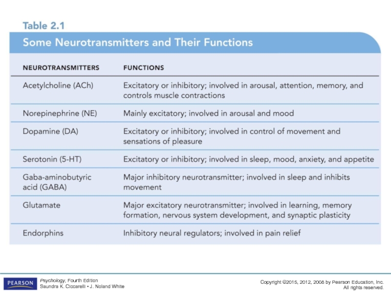
LO 2.2 How Neurons Use Neurotransmitters to Communicate

Слайд 14Neuron Communication
Synaptic vesicles: sack-like structures found inside the axon terminal containing
neurotransmitter: chemical found in the synaptic vesicles which, when released, has an effect on the next cell
LO 2.2 How Neurons Use Neurotransmitters to Communicate

Слайд 15Neuron Communication
synapse/synaptic gap: microscopic fluid-filled space between the rounded areas on
receptor sites: holes in the surface of the dendrites or certain cells of the muscles and glands, which are shaped to fit only certain neurotransmitters
LO 2.2 How Neurons Use Neurotransmitters to Communicate

Слайд 16
Figure 2.4 Reuptake of Dopamine
Dopamine is removed from the synapse by

Слайд 17Neuron Communication
Neurons must be turned ON and OFF
excitatory neurotransmitter: neurotransmitter that
inhibitory neurotransmitter: neurotransmitter that causes the receiving cell to stop firing
LO 2.2 How Neurons Use Neurotransmitters to Communicate

Слайд 18Neuron Communication
Chemical substances can affect neuronal communication
agonists: mimic or enhance the
antagonists: block or reduce a cell’s response to the action of other chemicals or neurotransmitters
LO 2.2 How Neurons Use Neurotransmitters to Communicate

Слайд 20Cleaning up the Synapse
reuptake: process by which neurotransmitters are taken back
enzyme: complex protein that is manufactured by cells
one enzyme specifically breaks up acetylcholine because muscle activity needs to happen rapidly; reuptake would be too slow
LO 2.2 How Neurons Use Neurotransmitters to Communicate


Слайд 22Central Nervous System
Central nervous system (CNS): part of the nervous system
spinal cord: a long bundle of neurons that carries messages to and from the body to the brain that is responsible for very fast, lifesaving reflexes
LO 2.3 How the Brain and Spinal Cord Interact
: part of the nervous system consisting of the brain")
Слайд 23The Reflex Arc: Three Types of Neurons
Sensory neuron: a neuron that
also called an afferent neuron
Motor neuron: a neuron that carries messages from the central nervous system to the muscles of the body
also called an efferent neuron
LO 2.3 How the Brain and Spinal Cord Interact

Слайд 24The Reflex Arc: Three Types of Neurons
Interneuron: a neuron found in
interneurons also make up the bulk of the neurons in the brain
LO 2.3 How the Brain and Spinal Cord Interact

Слайд 25The Reflex Arc: Three Types of Neurons
Neuroplasticity: the ability to constantly
LO 2.3 How the Brain and Spinal Cord Interact

Слайд 26Peripheral Nervous System
Peripheral nervous system (PNS): all nerves and neurons that
divided into the:
somatic nervous system
autonomic nervous system
LO 2.4 Somatic and Autonomic Nervous Systems
: all nerves and neurons that are not contained in")

Слайд 28Somatic Nervous System
Soma = “body”
Somatic nervous system: division of the PNS
sensory pathway: nerves coming from the sensory organs to the CNS consisting of sensory neurons
LO 2.4 Somatic and Autonomic Nervous Systems

Слайд 29Somatic Nervous System
Somatic nervous system (cont’d)
motor pathway: nerves coming from the
LO 2.4 Somatic and Autonomic Nervous Systems
motor pathway: nerves coming from the CNS to the voluntary")
Слайд 30Autonomic Nervous System
Autonomic nervous system (ANS)
division of the PNS consisting of
LO 2.4 Somatic and Autonomic Nervous Systems
division of the PNS consisting of nerves that control all")
Слайд 31Autonomic Nervous System
Autonomic Nervous System (ANS) (cont’d)
sympathetic division (fight-or-flight system): part
parasympathetic division: part of the ANS that restores the body to normal functioning after arousal and is responsible for the day-to-day functioning of the organs and glands
LO 2.4 Somatic and Autonomic Nervous Systems
 (cont’d)sympathetic division (fight-or-flight system): part of the ANS that")

Слайд 33The Endocrine Glands
Endocrine glands: glands that secrete chemicals called hormones directly
hormones: chemicals released into the bloodstream by endocrine glands
LO 2.5 How Hormones Interact with the Nervous System and Affect Behavior


Слайд 35The Endocrine Glands
pituitary gland: gland located in the brain that secretes
pineal gland: endocrine gland located near the base of the cerebrum that secretes melatonin
thyroid gland: endocrine gland found in the neck that regulates metabolism
pancreas: endocrine gland that controls the levels of sugar in the blood
LO 2.5 How Hormones Interact with the Nervous System and Affect Behavior

Слайд 36The Endocrine Glands
gonads: the sex glands; secrete hormones that regulate sexual
ovaries: the female gonads
testes: the male gonads
adrenal glands: endocrine glands located on top of each kidney
secrete over thirty different hormones to deal with stress, regulate salt intake
provide a secondary source of sex hormones affecting the sexual changes that occur during adolescence
LO 2.5 How Hormones Interact with the Nervous System and Affect Behavior

Слайд 37Looking inside the Living Brain
Clinical Studies
deep lesioning: insertion of a thin,
electrical stimulation of the brain (ESB): milder electrical current that causes neurons to react as if they had received a message
human brain damage
LO 2.6 Study of the Brain and How It Works

Слайд 38Looking inside the Living Brain
Clinical Studies
transcranial magnetic stimulation (TMS), magnetic pulses
repetitive TMS (rTMS).
transcranial direct current stimulation (tDCS),
human brain damage
LO 2.6 Study of the Brain and How It Works
, magnetic pulses are applied to the")
Слайд 39Mapping Structure
computed tomography (CT): brain-imaging method using computer-controlled X-rays of the
magnetic resonance imaging (MRI): brain-imaging method using radio waves and magnetic fields of the body to produce detailed images of the brain
LO 2.6 Study of the Brain and How It Works
: brain-imaging method using computer-controlled X-rays of the brainmagnetic resonance imaging (MRI):")
Слайд 40Mapping Structure
Mapping Function
electroencephalogram (EEG): records electric activity of the brain below
magnetoencephalography (MEG)
positron emission tomography (PET): radioactive sugar is injected into the subject and a computer compiles a color-coded image of brain activity of the brain; lighter colors indicate more activity
LO 2.6 Study of the Brain and How It Works
: records electric activity of the brain below specific areas of the")
Слайд 41Mapping Structure
Mapping Function (cont’d)
single photon emission computed tomography (SPECT): similar to
functional MRI (fMRI): a computer makes a sort of “movie” of changes in the activity of the brain using images from different time periods
LO 2.6 Study of the Brain and How It Works
single photon emission computed tomography (SPECT): similar to PET, but uses different")

Слайд 43The Hindbrain
The Hindbrain
medulla: first large swelling at the top of the
responsible for life-sustaining functions such as breathing, swallowing, and heart rate
pons: larger swelling above the medulla that connects the top of the brain to the bottom
plays a part in sleep, dreaming, left–right body coordination, and arousal
LO 2.7 Structures and Functions of the Bottom Part of Brain

Слайд 44The Hindbrain
reticular formation (RF): area of neurons running through the middle
responsible for selective attention
cerebellum: part of the lower brain located behind the pons
controls and coordinates involuntary, rapid, fine motor movement
LO 2.7 Structures and Functions of the Bottom Part of Brain
: area of neurons running through the middle of the medulla and")

Слайд 46Structures under the Cortex
Limbic system: a group of several brain structures
thalamus: part of the limbic system located in the center of the brain
relays sensory information from the lower part of the brain to the proper areas of the cortex
processes some sensory information before sending it to its proper area
LO 2.8 Structures that Control Emotion, Learning, Memory, and Motivation

Слайд 47Structures under the Cortex
Limbic System (cont’d)
hypothalamus: small structure in the brain
responsible for motivational behavior such as sleep, hunger, thirst, and sex
hippocampus: curved structure located within each temporal lobe
responsible for the formation of long-term memories and the storage of memory for location of objects
LO 2.8 Structures that Control Emotion, Learning, Memory, and Motivation
hypothalamus: small structure in the brain located below the thalamus")
Слайд 48Structures under the Cortex
Limbic System (cont’d)
amygdala: brain structure located near the
responsible for fear responses and the memory of fear
cingulate cortex: the limbic structure actually found in the cortex
plays important roles in cognitive and emotional processing
LO 2.8 Structures that Control Emotion, Learning, Memory, and Motivation
amygdala: brain structure located near the hippocampusresponsible for fear responses")
Слайд 49Cortex
cortex: outermost covering of the brain consisting of densely packed neurons
responsible
corticalization: wrinkling of the cortex
allows a much larger area of cortical cells to exist in the small space inside the skull
LO 2.9 Parts of Cortex Controlling Senses and Movement

Слайд 50Cerebral Hemispheres
cerebral hemispheres: the two sections of the cortex on the
corpus callosum: thick band of neurons that connects the right and left cerebral hemispheres
LO 2.9 Parts of Cortex Controlling Senses and Movement


Слайд 52Four Lobes of the Brain
occipital lobe: section of the brain located
primary visual cortex: processes visual information from the eyes
visual association cortex: identifies and makes sense of visual information
LO 2.9 Parts of Cortex Controlling Senses and Movement

Слайд 53Four Lobes of the Brain
parietal lobes
sections of the brain located at
somatosensory cortex: area of neurons running down the front of the parietal lobes
responsible for processing information from the skin and internal body receptors for touch, temperature, body position, and possibly taste
LO 2.9 Parts of Cortex Controlling Senses and Movement


Слайд 55Four Lobes of the Brain
temporal lobes: areas of the cortex located
primary auditory cortex: processes auditory information from the ears
auditory association cortex: identifies and makes sense of auditory information
LO 2.9 Parts of Cortex Controlling Senses and Movement

Слайд 56Four Lobes of the Brain
frontal lobes: areas of the cortex located
motor cortex: section of the frontal lobe located at the back; responsible for sending motor commands to the muscles of the somatic nervous system
LO 2.9 Parts of Cortex Controlling Senses and Movement

Слайд 57Association Areas of Cortex
association areas: areas within each lobe of the
LO 2.10 Parts of Cortex Responsible for Higher Thought

Слайд 58Association Areas of Cortex
Broca’s aphasia: condition resulting from damage to Broca’s
causes the affected person to be unable to speak fluently, to mispronounce words, and to speak haltingly
LO 2.10 Parts of Cortex Responsible for Higher Thought

Слайд 59Association Areas of Cortex
Wernicke’s aphasia: condition resulting from damage to Wernicke’s
causes the affected person to be unable to understand or produce meaningful language
LO 2.10 Parts of Cortex Responsible for Higher Thought

Слайд 60Association Areas of Cortex
spatial neglect: condition produced by damage to the
results in an inability to recognize objects or body parts in the left visual field
LO 2.10 Parts of Cortex Responsible for Higher Thought

Слайд 61Split-Brain Research
Cerebrum: the upper part of the brain consisting of the
LO 2.11 Differences between the Left and Right Sides of the Brain

Слайд 62Split-Brain Research
Split-Brain Research
study of patients with severed corpus callosum
involves sending messages
demonstrates right and left brain specialization
LO 2.11 Differences between the Left and Right Sides of the Brain


Слайд 64Results of Split-Brain Research
left side of the brain
seems to control language,
processes information sequentially, and enables one to speak
right side of the brain
controls emotional expression, spatial perception, recognition of faces, patterns, melodies, and emotions
it processes information globally and cannot influence speech
LO 2.11 Differences between the Left and Right Sides of the Brain

Слайд 65Attention-Deficit/Hyperactivity Disorder
Causes of ADHD have highlighted the likelihood of more than
Current research is looking at a variety of areas including environmental factors such as low-level lead exposure, genetic influences, the role of heredity and familial factors, and personality factors
LO 2.12 Some Potential Causes of Attention-Deficit/Hyperactivity Disorder