Слайд 1
LECTURE
PROTOZOA AS PARASITES
OF HUMAN BEING
ZAPOROZHYE STATE MEDICAL UNIVERSITY
DEPARTMENT OF
MEDICAL BIOLOGY
Composed by
Doctor of Philosophy
Popovich A. P.
madbio@zsmu.zp.ua
Zaporozhye - 2016
Слайд 2QUESTIONS
-General features
-Protozoa Taxonomy
-Characteristics of: Lobosea,
Zoomastigophora, Sporozoa, Litostomatea
Слайд 3General Features
1. Protozoa are microscopic, unicellular animals whose single cell performs
all activities of the multicellular organism: nutrition. respiration, locomotion, growth, excretion, sensitivity and reproduction.
2. The protozoan body is bounded by a delicate plasma membrane, which does not maintain a definite shape (ex. Amoeba). The shape of the body is maintained by the pellicle which is a double membrane and may be regarded as an exoskeleton (Ex. Euglena, Paramecium).
3. Beneath the pellicle is a peripheral ectoplasm - clear, dense and firm. Below the ectoplasm is a semi fluid, granular endoplasm with fat, glycogen and nucleus (one or two).
Слайд 44. Some vacuoles can be found in the endoplasm:
- food
vacuoles – for digestion and
- contractile vacuoles – for osmoregulation and for elimination of waste products, some dissolved CO2 , and the excess of water
5. Respiration may be effected either aerobically by oxidation or anaerobically, by splitting up of complex chemical substances into simple compounds.
6. Locomotion either by:
- producing pseudopodia (Amoeba)
- using flagella (Euglena, Trypanosoma) or cilia (Paramecium)
7. Protozoa reproduce:
- asexually by binary fission or
- sexually by conjugation
8. In unfavorable periods they from cysts. The cysts also provide means of dispresal and thus colonization of fresh territory.
Слайд 5PROTOZOA TAXONOMY
CLASS
LOBOSEA
Class
Zoomastigophora
Class
Litostomatea
Class
Sporozoa
Entamoeba
histolitica
Entamoeba
coli
Trypanosoma
gambiense
Trypanosoma
cruzi
Plasmodium
vivax
P.malariae
Paramecium
Caudatum
Balantidium
coli
Слайд 6Some of the most important disease – producing Protozoa are:
Entamoeba, Leishmania, Trypanosoma,
Lamblia, Toxoplasma, Plasmodium, Balantidium.
Слайд 8Entamoeba Histolitica
The fully grown form (trophozoite) is less or more rounded
with outer clear ectoplasme and urner granular endoplasme. It has a large round nucleus.
Ent. Histolitica can be found in forms:
- forma magna-pathogenic,
- forma minuta-the main form of existence,
- cyst form-invasive stage.
Слайд 9Entamoeba Histolitica – forma minuta is an unpathogenic commensal. It inhabits
the cavity of the large intestine, feeds on bacteria, detritous, reproduces, turns into cysts which passed out with the faeces.
Forma minuta changes into forma magna (pathogenic form) which penetrates into the cells of intestine causes disease amoebic dysentery or amoebiasis. In chronic amoebiasis the parasites bore into the blood vessels and are carried to the liver. In the liver the parasites produce abscesses.
Invasion of a person takes place when he swallows cysts with water and food.
Слайд 10Trypanosomes.
They are parasites in the blood, lymph and tissues.
The
adult form has a fusiform body pointed at both ends and covered with a firm pellicle. A longthread-like axoneme is joined to the cell by an undulating membrane and is continued along and beyond the body as a flagellum. There is a large nucleus in the middle of the cell.
Слайд 11 Three species are parasitic in man:
-Trypanosoma gambiense,
-Trypanosoma rhodesiense,
-Trypanosoma cruzi.
Tr. Gambiense and Tr. rhodesiense cause the sleeping-sickness (fever,anemia,enlargment of lymphatic nodes,inflammation of brain and cerebral membranes) in man in Africa. They are found in the blood of antilops from where they are transmitted to humans by tse-tse flies.
Tr. Cruzi is spread in South and Central America.It is transmitted to humans by bugs “Triatoma megista” and causes chagas disease (American Trypanosomiasis). Natural resevoirs are rats, squrrels, dogs, opossumes.
Слайд 13Leishmania.
It is pathogen of leishmaniasis (cutaneous or visceral).
In infected tissues (cells) Leishmania parasites occur as spherical or oval organisms, non-flagellated about 2-6 mm in length.The cytoplasm contains one or several vacuoles, a spherical nucleus and blepharoplast.
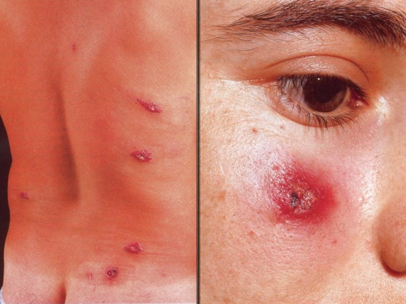
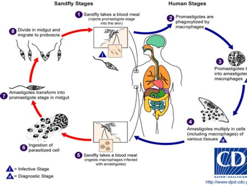
Leishmania tropica is the causative agent of cutaneous leishmaniasis which is transmitted to humans by sand-flies of Phlebotomus genus.
At the place of sand-flie’s sting a red papular appears and then ulcer forms. The ulcer cicatrizes and forms a scar that remains after treatment.
Слайд 15 Leismania domovani causes visceral leishmaniosis (or Kala-azar). The parasite
attacks blood vessels, lymphatics, spleen, liver, red bone marrow. The disease manifestes itself by fever, enlargment of the spleen, liver, by rheumatic pains.
Слайд 20Lamblia (Giardia).
It is an agent of lambliasis. The adult form
(trophozoite) has a pear shape, four pairs of flagella, two nuclei, sucking discs and a supporting shift-axostyle.
The parasite forms oval-shaped cysts, which are invasional stage. It locates in the small intestine and breaks down parental digestion. Human being invaded by swallowing cysts with water and food.
Слайд 22Trichomonas vaginalis.
Trichomonas vaginalis is an agent of uro-genital trichomoniasis. It
causes inflammation of mucose membrane of urethra, vagina.
The invasion comes upon sexual contacts and by using the things of personal hygiene (sponges, towels), non-sterilized medical instruments.
Слайд 24Toxoplasma gondii.
It causes toxoplasmosis and can be found in the
forms:
- adult forms-trophozoites,
- cysts.
Trophozoites are intra-cellular parasites about 6×2 mm, crescent-shaped with central nucleus. They multiply by binary fission and form pseudocysts .Pseudocysts are intra-cellular collection of trophozoites in cells. The cell wall of pseudocyst is a very thin membrane, that can be easyly distroyed when parasites grow.
Слайд 25 Protecting themselves from host’s immune system the parasites forms thick
capsules with a lot of trophozoites inside.
These capsules are known as a true cysts.
Oocysts develop in final host (cat) during sexual reproduction. Each oocyst contains two sporocysts with 4 sporozoites and found in stool of infected cats.
Cats are the final hosts of Toxoplasma which reproduces sexually in these animals. Humans, birds in which parasite reproduces asexually are intermediate hosts.
Слайд 26
Humans ingest oocysts with meat , milk and dairy products
of animals sick with toxoplasmosis, uncooked infected eggs and contaminated water or due to the direct contact with a cat.
Very dangerous is transplacental infection. It may cause embryo death or birth of a cripped child. In this case trophozoite (endozoit) is invasional stage.
Infection is often asymptomatic.
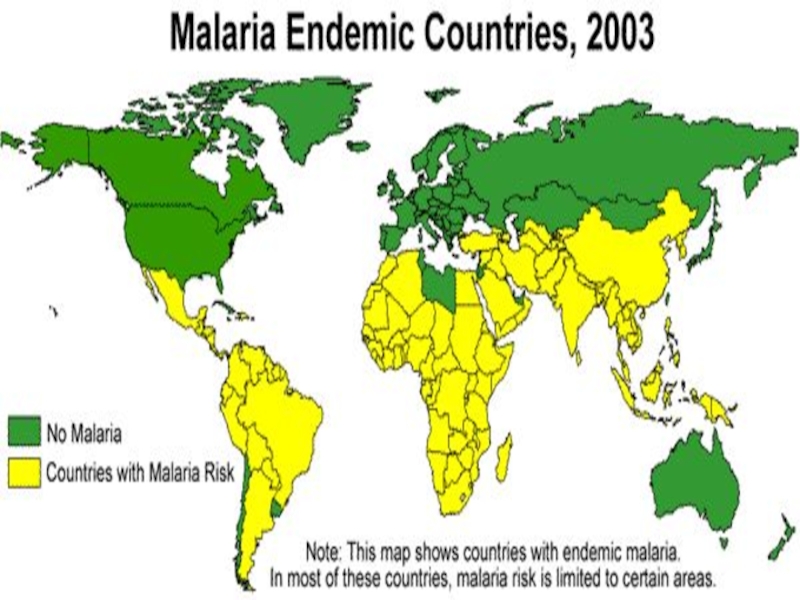
Слайд 28Plasmodium.
The malarial parasites of man include four species:
- Pl.
vivax - agent of tertian malaria,
- Pl. malaria - agent of quartan malaria,
- Pl.falciparum - agent of tropical malaria,
- Pl.ovale – agent of tertien ovale -malaria.
Слайд 29
Life cycle involves sexual stage (sporogony) in the
mosquito Anopheles and asexual stage (schizogony) in man. Man is an intermediate host and mosquito is a definitive host.
The life cycle passes 3 stages:
a) in man: - Exo - erythrocytic schizogony (liver phase),
- Erythrocytic schizogony (blood phase).
b) in mosquito:
- Sporogony.
Clinical features of malaria include series of febrile paroxysms followed by anemia and splenomegaly.
Слайд 32Balantidium coli.
Balantidium coli is the only infusorial parasite of man
which causes the disease balantidiasis. It has two stages:
- a trophozoite stage,
- a cyst stage.
The parasite has an asymmetrical oval body covered with cilia. Its anterior end is more pointed than the posterior and has the opening, knowing as peristome which leads to the cytostome (mouth) and then into a short cytopharynx.
Слайд 33 The posterior end of the body has an anal
pore. The parasite has two nuclei: the macronucleus and the micronucleus, food contractile vacuole. It multiplies by longitudinal fission and encysts.
Infection occurs when the cysts are ingested through contaminated food or water.
B. coli lives in the caecum and colon of humans, pigs, rats and other mammals.
It causes ulceration of the large intestine, diarrhea which may become chronic.


 is less or more rounded with outer clear ectoplasme")


. In infected tissues (cells) Leishmania parasites")
. The parasite attacks blood vessels, lymphatics, spleen,")
. It is an agent of lambliasis. The adult form (trophozoite) has a pear")


 in the mosquito Anopheles and asexual stage")