- Главная
- Разное
- Дизайн
- Бизнес и предпринимательство
- Аналитика
- Образование
- Развлечения
- Красота и здоровье
- Финансы
- Государство
- Путешествия
- Спорт
- Недвижимость
- Армия
- Графика
- Культурология
- Еда и кулинария
- Лингвистика
- Английский язык
- Астрономия
- Алгебра
- Биология
- География
- Детские презентации
- Информатика
- История
- Литература
- Маркетинг
- Математика
- Медицина
- Менеджмент
- Музыка
- МХК
- Немецкий язык
- ОБЖ
- Обществознание
- Окружающий мир
- Педагогика
- Русский язык
- Технология
- Физика
- Философия
- Химия
- Шаблоны, картинки для презентаций
- Экология
- Экономика
- Юриспруденция
Метаботропные рецепторы. Подсемейство рецепторов презентация
Содержание
- 1. Метаботропные рецепторы. Подсемейство рецепторов
- 2. Родопсин-подобные метаботропные рецепторы (выделены синим)
- 3. Ацетилхолиновые метаботропные рецепторы Агонисты: АцХ, карбахол, мускарин
- 4. Ацетилхолиновые метаботропные рецепторы Аmino acid sequence and
- 5. Ацетилхолиновые метаботропные (мускариновые) рецепторы
- 6. Агонисты (выделены)
- 7. Мускариновые рецепторы в парасимпатической системе
- 8. Ацетилхолиновые метаботропные рецепторы
- 9. Ацетилхолиновые метаботропные рецепторы Выделено 5 подтипов мускариновых рецепторов - М(1-5)
- 10. Ацетилхолиновые метаботропные рецепторы Выделено 5 подтипов мускариновых рецепторов - мАцХР(1-5)
- 11. Примеры эффектов мускариновых рецепторов М1 (М3, М5)
- 12. Примеры эффектов мускариновых рецепторов M1 receptor This
- 13. Примеры эффектов мускариновых рецепторов M3 receptor The
- 14. Примеры эффектов мускариновых рецепторов M4 receptor M4
- 15. Общая схема каскадов, инициируемых мускариновыми рецепторами
- 16. Каскады, инициируемые мускариновыми рецепторами М1,
- 17. GIRK - G protein-coupled inwardly-rectifying potassium channel
- 18. M1, M3 and M5 mAChRs preferentially couple
- 19. Ацетилхолиновые метаботропные рецепторы: эффекты активации The M1,
- 20. Каскад m1, вызывающий снижение К +-тока из-за
- 21. m1: Снижение К+-проводимости происходит в результате фосфорилирования субъединиц канала с участием PKC
- 22. m1: Снижение К+-проводимости приводит к снижению выходящего
- 23. Активация М1-рецептора сопровождается снижением флуоресценции (светимости) специального
- 24. Ацетилхолиновые метаботропные рецепторы: эффекты активации One of
- 25. Generalized diagram of G protein-gated ion channel:
- 26. GIRK - G protein-coupled inwardly-rectifying potassium channel (усиление входящего К+-тока)
- 27. Протон-активируемые метаботропные рецепторы из класса родопсин-подобных рецепторов
- 28. Протон-активируемые метаботропные рецепторы (каскады) Signaling mechanisms of
- 29. Протон-активируемые метаботропные рецепторы (каскады, предполагаемая роль OGR1 в секреции инсулина и его синтезе в β-клетках)
- 30. Glucose is transported into the β-cell by
- 31. Глютаматные метаботропные рецепторы (выделены оранжевым)
- 32. Глютаматные метаботропные рецепторы
- 33. Глютаматные метаботропные рецепторы включают восемь типов метаботропных
- 34. Метаботропные глутаматные рецепторы The mGluRs
- 35. Каскады групп I и II глутаматных метаботропных рецепторов
- 36. Метаботропные глутаматные рецепторы Group I
- 37. Метаботропные глутаматные рецепторы Group II & Group
- 38. Вкусовые рецепторы T1R - обеспечивает вкус «сладкого»
- 39. mGluR обеспечивают рецепцию вкуса аминокислот (в т.ч.
- 40. Вкусовые рецепторы A model for the major
- 41. Transient receptor potential (TRP) channel
- 42. Bitter, sweet & umami taste transduction
- 43. Вкусовые рецепторы
- 44. Пример действия mGluR6 в биполярных клетках сетчатки
- 45. Темновой ток в фоторецепторах сетчатки - деполяризация
- 46. Темновой ток в фоторецепторах сетчатки на свету устраняется
- 47. Сигнал от фоторецепторов на On- и OFF-биполярных клетках сетчатки
- 48. mGluR6 в биполярных клетках. Раньше полагали так:
- 49. В темноте (при деполяризации) фоторецепторы выделяют Glu
- 50. В темноте (при деполяризации) фоторецепторы выделяют Glu
- 51. В темноте (при деполяризации) фоторецепторы выделяют Glu
- 52. mGluR6 в биполярных клетках. Роль TRPM1-каналов В
- 53. Метаботропные ГАМКВ рецепторы Широко распространены в ЦНС
- 54. Тормозное действие осуществляется через Gi/o-белки:
- 55. Внеклеточно Са2+-активируемые метаботропные рецепторы
- 56. Внеклеточно Са2+-активируемые метаботропные рецепторы (модель димерной формы)
- 57. Внеклеточно Са2+-активируемые метаботропные рецепторы (каскады, активируются множеством лигандов)
- 58. Рецепторы катехоламинов
- 59. Адренергические рецепторы
- 60. Адренергические рецепторы
- 61. Адренергические рецепторы (α-тип)
- 62. Адренергические рецепторы (β -тип)
- 63. Каскады адренергических рецепторов Gq Gi Gs
- 64. Каскады адренергических рецепторов Adrenaline or noradrenaline
- 65. Адренергические рецепторы группа α1 сопряжена с Gq-белком
- 66. Адренергические рецепторы группа α2 сопряжена с Gi/Go-белками
- 67. Адренергические рецепторы группа β сопряжена с Gs-белком activate adenylyl cyclase activity
- 68. Activate adenylyl cyclase activity
- 69. Классические и неклассические каскады β-рецепторов
- 70. Классические и неклассические каскады β-рецепторов
- 71. Дофаминовые рецепторы
- 72. Дофаминовые рецепторы: каскады подразделяют на два семейства:
- 73. Дофаминовые рецепторы D1-семейства Activation of D1-like family
- 74. Дофаминовые рецепторы D2-семейства D2-like activation is coupled
- 75. Дофаминовые рецепторы D2-семейства D2-like activation is coupled
- 76. Дофаминовые рецепторы D2-семейства D3 Maximum expression of
- 77. Дофаминовые рецепторы: общая характеристика
- 78. Дофаминовые рецепторы: функции
- 79. Серотониновые рецепторы
- 80. Серотониновые рецепторы
- 81. Серотониновые рецепторы (каскады) Mammalian 5-HT receptor subtypes
- 82. Серотониновые рецепторы (каскады) 5-HTR signaling pathways and
- 83. Серотониновые рецепторы (каскады)
- 84. Серотониновые рецепторы (каскады)
- 85. Серотониновые рецепторы: функции
- 86. Гистаминовые рецепторы
- 87. Каскады гистаминовых рецепторов
- 88. Гистаминовые рецепторы
- 89. Гистаминовые рецепторы H1 рецептор через Gq-белки активирует
- 90. Пуриновые метаботропные рецепторы: АТФ Метаботропные Р-рецепторы –
- 91. Пуриновые метаботропные рецепторы: АТФ
- 92. Пуриновые метаботропные рецепторы: АТФ
- 93. Аденозиновые рецепторы
- 94. Аденозиновые рецепторы
- 95. Пуриновые метаботропные рецепторы: аденозиновые Через пресинаптические А1-рецепторы
- 96. Обонятельные рецепторы
- 97. trace-amine-associated receptors
- 98. Обонятельные рецепторы A model for the transduction
- 99. Обонятельные рецепторы A model for chemosensory transduction
- 102. Опиоидные рецепторы μ-, δ-, κ- и ORL(opioid
- 103. Опиоидные рецепторы
- 104. Inhibition of pain: - endorphins
- 105. Тахикинины Включают вещество Р (SP), нейрокинин А
- 106. Функциональная роль некоторых пептидов
- 107. Эндоканнабиноиды Анандамид (anandamide)
- 108. Рецепторы эндоканнабиноидов Идентифицировано два эндоканнабиноидных рецептора –
- 109. Рецепторы эндоканнабиноидов Идентифицировано два эндоканнабиноидных рецептора –
- 110. Рецепторы эндоканнабиноидов Пресинаптические СВ1 снижают высвобождение глютамата,

")
Слайд 3Ацетилхолиновые метаботропные рецепторы
Агонисты: АцХ, карбахол, мускарин
Антагонисты: атропин, скополамин, галламин
мАцХР локализованы
в
которые иннервируются постганглионарными волокнами вегетативной НС (парасимпатический отдел);
в пресинаптических мембранах постганглионарных нейронов симпатической НС (торможение со стороны постганглионарных нейронов парасимпатической НС );
в пресинаптических мембранах мотонейронов в нервно-мышечных
синапсах;
- в ЦНС в пост- и пресинаптических мембранах.

Слайд 4Ацетилхолиновые метаботропные рецепторы
Аmino acid sequence and transmembrane domain structure of the
Amino acids that are identical among the m1, m2, m3 and m4 receptors are dark orange. The shaded cloud represents the approximate region that determines receptor–G-protein coupling. Arrows denote amino acids important for specifying G protein coupling. Amino acids predicted to be involved in agonist or antagonist binding are denoted by white letters.
55–70 kDa

 рецепторы")
")


")
Слайд 10Ацетилхолиновые метаботропные рецепторы
Выделено 5 подтипов мускариновых рецепторов - мАцХР(1-5)
")
Слайд 11Примеры эффектов мускариновых рецепторов
М1 (М3, М5) в вегетативных ганглиях и в
ИФ3 повышает концентрацию Са2+ в цитозоле, который вместе с
ДАГ активирует протенкиназу С.
М2 (М4) в ЦНС и сердце активируют Gi-белки и подавляют аденилатциклазу, уменьшая концентрацию цАМФ, Go-белки (βγ-димер) увеличивают К+- и снижают Са2+-проводимость
 в вегетативных ганглиях и в желудке через Gq-белки активируют")
Слайд 12Примеры эффектов мускариновых рецепторов
M1 receptor
This receptor is found mediating slow EPSP
It is predominantly found bound to G proteins of class Gq which use upregulation of phospholipase C and therefore inositol trisphosphate and intracellular calcium as a signalling pathway. However, Gi (causing a downstream decrease in cAMP) and Gs (causing an increase in cAMP) have also been shown to be involved in interactions in certain tissues.
M2 receptor
The M2 muscarinic receptors are located in the heart, where they act to slow the heart rate down to normal sinus rhythm after stimulatory actions of the parasympathetic nervous system, by slowing the speed of depolarization. They also reduce contractile forces of the atrial cardiac muscle, and reduce conduction velocity of the atrioventricular node (AV node). It also serves to slightly decrease the contractile forces of the ventricular muscle.
M2 muscarinic receptors act via a Gi type receptor, which causes a decrease in cAMP in the cell, generally leading to inhibitory-type effects. Effects include formation of IP3 and DAG.

Слайд 13Примеры эффектов мускариновых рецепторов
M3 receptor
The M3 muscarinic receptors are located at
The M3 receptors are also located in many glands which help to stimulate secretion in salivary glands and other glands of the body.
Like the M1 muscarinic receptor, M3 receptors are G proteins of class Gq which upregulate phospholipase C and therefore inositol trisphosphate and intracellular calcium as a signalling pathway.

Слайд 14Примеры эффектов мускариновых рецепторов
M4 receptor
M4 receptors are found in the CNS.
M5 receptor
Location of M5 receptors is not well known. Like the M1 and M3 muscarinic receptor, M5 receptors are coupled with G proteins of class Gq which upregulate phospholipase C and therefore inositol trisphosphate and intracellular calcium as a signalling pathway.


Слайд 16Каскады, инициируемые мускариновыми рецепторами
М1, М3 и М5 структурно похожи, активируют фосфолипазу
М1 также активирует гуанилатциклазу (Gs) и ингибирует аденилатциклазу (Gi)
М2 и М4 структурно похожи, ингибируют (Gi) аденилатциклазу открывают К+-каналы, закрывают Са2+-каналы (Go).
 М1 также")

Слайд 18M1, M3 and M5 mAChRs preferentially couple to G-proteins of the
whereas M2 and M4 receptors typically activate G-proteins of the Gi/Go family.
Agonist occupancy of the two groups of mAChRs results in the activation of different downstream effector proteins, as indicated, although some effectors (e.g., mitogen-activated protein kinase) (MAPK) are activated by both groups of receptors. Note that the effects of mAChR activation are mediated by both the α and βγ subunits of the G-proteins. An increase or decrease in the activity of the effector mechanism is indicated by the direction of the arrow. GIRK, G-protein–activated inwardly rectifying K+ channel; PLCβ, phosphoinositide-specific phospholipase C.
Muscarinic cholinergic receptors can be subdivided based upon their G-protein–coupling characteristics and effector mechanisms

Слайд 19Ацетилхолиновые метаботропные рецепторы:
эффекты активации
The M1, M3 and M5 mAChRs preferentially couple
These second messengers are responsible for the mobilization of intracellular Ca2+ and activation of protein kinase C (PKC) and subsequently, that of mitogen-activated protein kinase (MAPK).
M1 receptors have also been shown to inhibit a voltage-sensitive current known as M-current (“M” for muscarinic). mAChR–mediated inhibition of K+ efflux through the M-channels results in the slow depolarization of the cell and a facilitation of repetitive cell firing.

Слайд 20Каскад m1, вызывающий снижение К +-тока из-за блокады К +-проводимости (IM-ток)
(PKC
(PKC – активируется DAG и Ca2+)")
Слайд 21
m1: Снижение К+-проводимости происходит в результате фосфорилирования субъединиц канала с участием

Слайд 22m1: Снижение К+-проводимости приводит к снижению выходящего К+-тока (сравни а и
Oxo-M - muscarinic agonist
oxotremorine-methiodide
Выходящий ток
Спайковая активность
 и, как следствие,")
Слайд 23Активация М1-рецептора сопровождается снижением флуоресценции (светимости) специального красителя при его связывании
 специального красителя при его связывании с ИФ3. Снижение флуоресценции")
Слайд 24Ацетилхолиновые метаботропные рецепторы:
эффекты активации
One of the major consequences of the activation
M2 and M4 mAChRs can also cause a rapid activation of G-protein-coupled, inwardly rectifying K+-channels (GIRKs). However, activation of these channels, which results in membrane hyperpolarization, is a result of the direct interaction of the βγ subunits with the channel itself; no second messenger formation is required.
M2 and M4 receptors can also negatively modulate Ca2+ currents whereas they activate MAPK.

Слайд 25Generalized diagram of G protein-gated ion channel:
Typically, the activated effector protein
(B) The GTP-bound α-subunit in some cases can directly activate the ion channel.
(C) In other cases, the activated βγ-complex of the G protein may interact with the ion channel.
GIRK - G protein-coupled inwardly-rectifying potassium channel
активируют К+-каналы внутреннего выпрямления (К+-ток внутрь клетки)

")

Слайд 28Протон-активируемые метаботропные рецепторы
(каскады)
Signaling mechanisms of proton-sensing GPCRs:
- OGR1 is coupled with
- the proton-sensing role of G2A is in question.
Signaling mechanisms of proton-sensing GPCRs:- OGR1 is coupled with Gq/11 proteins and phospholipase")
Слайд 29Протон-активируемые метаботропные рецепторы
(каскады, предполагаемая роль OGR1 в секреции инсулина и его
")
Слайд 30Glucose is transported into the β-cell by the glucose transporter 2
http://www.nature.com/nature/journal/v414/n6865/fig_tab/414788a_F1.html
. By catalysing")
")

Слайд 33Глютаматные метаботропные рецепторы
включают восемь типов метаботропных рецепторов (mGluR), Са2+-чувствительные рецепторы и
Характеризуются длинными N- и С-терминалями. Лиганд-связывающий участок у mGluR локализован на N-терминалях двух субъединиц рецептора, которые связаны между собой дисульфидным мостиком.
Два цистеиновых остатка на внеклеточных петлях образуют дисульфидный мостик. Уникальной особенностью этого семейства рецепторов является короткая и высоко консервативная внутриклеточная петля ТМ5-ТМ6.
Агонист
, Са2+-чувствительные рецепторы и ГАМКВ рецепторы.Характеризуются длинными N-")
Слайд 34Метаботропные глутаматные рецепторы
The mGluRs perform a variety of functions in the
Подразделяются на три группы в соответствии со структурой и физиологической активностью. Внутри групп гомология составляет 60-70%, между группами – около 40%.


Слайд 36Метаботропные глутаматные рецепторы
Group I
The mGluRs in group I, including mGluR1
These receptors are also associated with Na+- and K+-channels. Their action can be excitatory, increasing conductance, causing more glutamate to be released from the presynaptic cell, but they also increase inhibitory postsynaptic potentials, or IPSPs. They can also inhibit glutamate release and can modulate voltage-dependent calcium channels.
Group I mGluRs, but not other groups, are activated by 3,5-dihydroxyphenylglycine (DHPG), a fact which is useful to experimenters because it allows them to isolate and identify them.

Слайд 37Метаботропные глутаматные рецепторы
Group II & Group III
The receptors in group II,
The chemicals 2-(2,3-dicarboxycyclopropyl)glycine (DCG-IV) and eglumegad activate only group II mGluRs, while 2-amino-4-phosphonobutyrate (L-AP4) activates only group III mGluRs. Several subtype-selective positive allosteric modulators have also now been developed which activate only the mGlu2 subtype, such as Biphenylindanone A.
LY-341,495 is a drug which acts as a selective antagonist blocking both of the group II metabotropic glutamate receptors, mGluR2 and mGluR3.

Слайд 38Вкусовые рецепторы
T1R - обеспечивает вкус «сладкого»
T2R/TRB - обеспечивает вкус «горького»
taste-mGluR4- обеспечивает вкус
")
Слайд 39mGluR обеспечивают рецепцию вкуса аминокислот
(в т.ч. глутамата, Umami)
Evidence also suggests that
Семейство TRP-каналов насчитывает 33 разновидности, разделенные на 8 подсемейств.
TRPM5- (transient receptor potential melastatin) каналы относятся к потенциал-зависимым каналам и активируются Са2+.
TRPM4 and TRPM5 form Ca2+-activated Na+ channels, impermeable for Ca2+.
!!! на рис. ошибка
Evidence also suggests that mGLuRs or taste")
Слайд 40Вкусовые рецепторы
A model for the major signaling mechanisms for the transduction
The individual steps are detailed in the text. Note that stimuli of each of these taste qualities interact with GPCRs: bitter stimuli with T2Rs, and sweet and umami stimuli with T1Rs. α-Gustducin has been implicated in the transduction of all three types of stimuli, but other α-subunits likely also couple to T1Rs or T2Rs in some TRC populations. PLC-β2 and the Ca2+-activated TRP channel subunit TRPM5 are essential for normal sweet, bitter and umami taste. The role of IP3 and the IP3R in the stimulus-dependent increase in intracellular Ca2+ as depicted are speculative.

 channel")

Слайд 43Вкусовые рецепторы
выделяется глюкагоно-
подобный пептид


Слайд 48mGluR6 в биполярных клетках. Раньше полагали так:
В темноте (при деполяризации)
На свету (при гиперполяризации) фоторецепторы перестают выделять Glu (фосфодиэстераза не активируется), и цГМФ-зависимый Na+/Са2+-ток восстанавливается, что приводит к деполяризации ON-биполяров.
 фоторецепторы выделяют Glu и")
Слайд 49В темноте (при деполяризации) фоторецепторы выделяют Glu и активируют mGluR6-рецепторы (R),
mGluR6 в биполярных клетках. Раньше полагали так:
 фоторецепторы выделяют Glu и активируют mGluR6-рецепторы (R), которые (Gi-белки) активируют фермент")
Слайд 50В темноте (при деполяризации) фоторецепторы выделяют Glu и активируют mGluR6-рецепторы, которые
mGluR6 в биполярных клетках. Современные представления:
?
?
Shen Y, Heimel JA, Kamermans M, et al. 2009. A transient receptor potential-like channel mediates synaptic transmission in rod bipolar cells.
J Neurosci 29: 6088–93.
Koike C, Sanuki R, Miyata K, et al. 2007. The functional analysis of TRPM1 in retinal bipolar cells.
Neurosci Res 58S: S41.
 фоторецепторы выделяют Glu и активируют mGluR6-рецепторы, которые через Go-белки активируют неизвестный")
Слайд 51В темноте (при деполяризации) фоторецепторы выделяют Glu и активируют mGluR6-рецепторы, которые
mGluR6 в биполярных клетках. Современные представления:
Shen Y, Heimel JA, Kamermans M, et al. 2009. A transient receptor potential-like channel mediates synaptic transmission in rod bipolar cells.
J Neurosci 29: 6088–93.
Koike C, Sanuki R, Miyata K, et al. 2007. The functional analysis of TRPM1 in retinal bipolar cells.
Neurosci Res 58S: S41.
 фоторецепторы выделяют Glu и активируют mGluR6-рецепторы, которые через Go-белки активируют неизвестный")
Слайд 52mGluR6 в биполярных клетках. Роль TRPM1-каналов
В отсутствие TRPM1-каналов (у мышей-нокаутов) на
реакции
палочковых ON-биполяров колбочковых ON-биполяров
В отсутствие TRPM1-каналов на свет
OFF-биполяры тормозятся (выходящие токи)
 на свет ON-биполяры не активируются")
Слайд 53Метаботропные ГАМКВ рецепторы
Широко распространены в ЦНС и вегетативной НС.
Тормозное действие осуществляется
αi-субъединица ингибирует аденилатциклазу;
β/γ-димер (Go) напрямую активирует К+-каналы;
β/γ-димер (Go) напрямую ингибирует Са2+-каналы.
ГАМКB рецептор гетеродимер и образован двумя субъединицами GABABR1 и GABABR2.
Агонист: (R)-baclofen
Антагонист: phaclofen

Слайд 54Тормозное действие осуществляется через Gi/o-белки:
αi-субъединица ингибирует аденилатциклазу;
β/γ-димер (Go)
β/γ-димер (Go) напрямую ингибирует Са2+-каналы.
ГАМКB рецептор гетеродимер и образован двумя субъединицами GABABR1 и GABABR2.
ГАМК связывается только с GABABR1-субъединицей
GABABR2 обеспечивает лишь аллостерическую модуляцию
Сигнализация осуществляется GABABR2-субъединицей через Gi/о-белки
 напрямую активирует К+-каналы; β/γ-димер (Go)")

")
Слайд 57Внеклеточно Са2+-активируемые метаботропные рецепторы
(каскады, активируются множеством лигандов)
")


")
")

Слайд 64Каскады адренергических рецепторов
Adrenaline or noradrenaline are receptor ligands to either
α1 couples to Gq, which results in incerased intracellular Ca2+ which results in e.g. smooth muscle contraction.
α2, on the other hand, couples to Gi, which causes a decrease of cAMP activity, resulting in e.g. smooth muscle contraction.
β receptors couple to Gs, and increases cAMP activity, resulting in e.g. heart muscle contraction, smooth muscle relaxation and glycogenolysis

Слайд 65Адренергические рецепторы группа α1 сопряжена с Gq-белком activates phospholipase C, leading to increased

Слайд 66Адренергические рецепторы
группа α2 сопряжена с Gi/Go-белками
inhibit adenylyl cyclase and stimulate phospholipase
activation of α2-adrenergic receptors leads to release of Gβγ resulting in activation of K+ channels and inhibition of Ca2+ channels.


Слайд 72Дофаминовые рецепторы: каскады
подразделяют на два семейства:
D1-like family (excitatory) D1 D5
 D1 D5 D2-like")
Слайд 73Дофаминовые рецепторы D1-семейства
Activation of D1-like family receptors (D1 и D5) is
 is coupled to the G")
Слайд 74Дофаминовые рецепторы D2-семейства
D2-like activation is coupled to the G protein Gαi,
D2-like activation is coupled also to the G protein Go,
которые активируют К+-каналы и инактивируют Са2+-каналы, обеспечивая тормозные процессы.

Слайд 75Дофаминовые рецепторы D2-семейства
D2-like activation is coupled to the G protein Gαi,
D2
There is a short version of D2 (D2Sh) and a long version of D2 (D2Lh):
The D2Sh are pre-synaptic situated, having modulatory functions (called autoreceptor, they regulate the neurotransmission by feed-back mechanisms, i.e., synthesis, storage and release of dopamine into the synaptic cleft).
The D2Lh may have the classic function of a post-synaptic receptor, i.e., keep going on the neurotransmission (excitatory or inhibitory) once blocked by a receptor antagonist or stimulated by the endogenous neurotransmitter itself or a synthetic full or partial agonist.

Слайд 76Дофаминовые рецепторы D2-семейства
D3
Maximum expression of dopamine D3 receptors is noted in
D4
The D4 receptor has the following variants D4.2, D4.3a, D4.3b, D4.4a, D4.4b, D4.4c, D4.4d, D4.4e, D4.5a, D4.5b, D4.6a, D4.6b, D4.7a, D4.7b, D4.7c, D4.7d, D4.8, D4.10.
These variants differ in a variable number tandem repeat domain present within the coding sequence of exon 3.
Some of these alleles are associated with greater incidence of certain diseases. For example, the D4.7 alleles have an established association with attention-deficit hyperactivity disorder.


Слайд 81Серотониновые рецепторы (каскады)
Mammalian 5-HT receptor subtypes and their signal transduction pathways.
The
AC, adenylyl cyclase;
cAMP, cyclic adenosine monophosphate;
GIRK, G protein-gated inwardly rectifying K+ channel;
ADPR, ADP-ribosyl cyclase;
cADPR, cyclic adenosine diphosphate ribose;
PLC, phospholipase C.
ADPR cADPR
Mammalian 5-HT receptor subtypes and their signal transduction pathways.The dotted lines show the")
Слайд 82Серотониновые рецепторы (каскады)
5-HTR signaling pathways and effectors.
Blue 5-HTR signal transduction in
PL phospholipase, ERK extracellular signal-regulated kinase, PK protein kinase, IP3 inositol triphosphate, DAG diacylglycerol, MAPK mitogen-activated protein kinase, NOS nitric oxide synthase, AHP after-hyperpolarization, JAK Janus kinase, STAT transcription pathway, ih hyperpolarization-activated current, Epa activated exchange factor.
5-HTR signaling pathways and effectors.Blue 5-HTR signal transduction in neurons, gray signaling linkages")
")
")


Слайд 89Гистаминовые рецепторы
H1 рецептор через Gq-белки активирует фосфолипазу С, вызывая синтез ИФ3.
H2 рецептор через Gs-белки активирует аденилатциклазу, вызывая увеличение Са2+-тока, что в конечном итоге приводит к возбудительным эффектам во внутренних органах (желудочно-кишечном тракте, в кровеносных и лимфатических сосудах).
H3 рецептор является ауторецептором и через Gi/o-белки напрямую снижает Са2+-проводимость, тем самым уменьшая выделение гистамина из пресинаптических окончаний (отрицательная обратная связь). H3 рецептор также описан как постсинаптический рецептор в стриатуме и коре мозга.
H4 инициирует хемотаксис тучных клеток и не задействован в цепях нейронной сигнализации.

Слайд 90Пуриновые метаботропные рецепторы: АТФ
Метаботропные Р-рецепторы – P2Y, P2T и P2U –
Семейство P2Y включает 12 метаботропных рецепторов, локализованных в постсинаптических мембранах.
Осуществляют свои эффекты главным образом через Gq-белки (активация фосфолипазы С), реже через Gi- и Gs-белки, соответственно, ингибируя и активируя аденилатциклазу.


Слайд 95Пуриновые метаботропные рецепторы: аденозиновые
Через пресинаптические А1-рецепторы (при сопряжении с Go-белками) аденозин
Активируя А2а-рецепторы, аденозин через Gs-белки активирует аденилатциклазу.
А2в-рецепторы через Gq-белки активируют фосфолипазу С.
В результате синтеза липидов в нейронах активируются Са2+-зависимые К+-каналы, что приводит к усилению следовой гиперполяризации и значительному тормозному эффекту на центральные нейроны.
Рецепторы А3 содержатся в нервной ткани в очень малом количестве, их функция в механизмах межнейронной сигнализации мало изучена. Предположительно они активируют фосфолипазу С.
 аденозин может уменьшать синаптическое выделение")


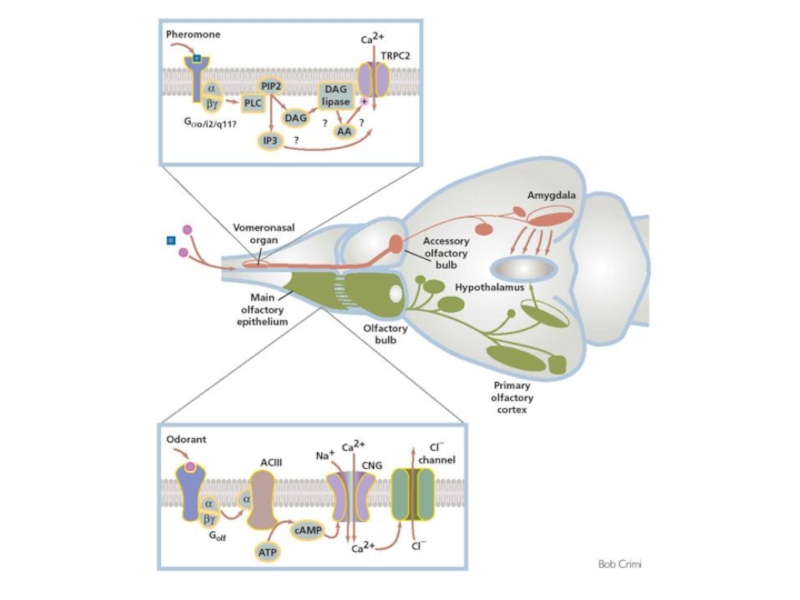
Слайд 98Обонятельные рецепторы
A model for the transduction of odors in canonical OSNs
The

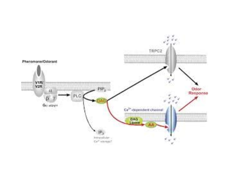
Слайд 99Обонятельные рецепторы
A model for chemosensory transduction in vomeronasal sensory neurons
The individual

Слайд 102Опиоидные рецепторы
μ-, δ-, κ- и ORL(opioid receptor-like)-рецепторы сопряжены с Gi/o-белками, которые
-рецепторы сопряжены с Gi/o-белками, которые обеспечивают закрытие Са2+-каналов (κ-рецепторы)")

Слайд 104Inhibition of pain:
- endorphins (enkephalins) = pain-inhibiting neurotransmitters produced by
descending fibers synapse (1) at the spinal cord dorsal horn release endorphins into synapse between sensory neurons (2) and ascending pain neurons (3)
endorphins have specific receptor sites on post-synaptic neurons
inhibitory action > opening of K+-channels
> closing Ca2+-channels
hyperpolarizing post-synaptic membrane act as pain killers by inhibiting pain signals along ascending pain neurons
1
2
3
 = pain-inhibiting neurotransmitters produced by reticular formation in braindescending")
Слайд 105Тахикинины
Включают вещество Р (SP), нейрокинин А (NKA) и нейрокинин В (NKB).
Афинность
Общий механизм сопряжен с Gq-белками, каскады фосфолипазы С (ИФ3/ДАГ). Эффект заключается в медленной деполяризации через закрытие К+-каналов.
, нейрокинин А (NKA) и нейрокинин В (NKB).Афинность лигандов (в порядке уменьшения)")

Слайд 107Эндоканнабиноиды
Анандамид (anandamide)
2-арахидонил-глицерол (2-arachidonylglycerol, 2-AG)
Синтезируются в результате повышения внутриклеточной концентрации Са2+. Механизм
Предполагается, что они диффундируют через клеточную мембрану и достигают соседних клеток.
Через свои рецепторы эндоканнабиноиды уменьшают выделение ГАМК из тормозных терминалей, предположительно действуя на потенциал-зависимые Са2+- и/или К+-каналы пресинаптической мембраны.
2-арахидонил-глицерол (2-arachidonylglycerol, 2-AG)Синтезируются в результате повышения внутриклеточной концентрации Са2+. Механизм высвобождения из клеток неизвестен.")
Слайд 108Рецепторы эндоканнабиноидов
Идентифицировано два эндоканнабиноидных рецептора – СВ1 и СВ2 (44% гомологии).
СВ1
Каскад с Gi/o-белками приводит к ингибированию аденилатциклазы и открытию Kir-каналов.
При активации Gβ/γ-белков пресинаптическими СВ1 блокируются Са2+-каналы.
.СВ1 сопряжен с Gi/o-белками (реже")
Слайд 109Рецепторы эндоканнабиноидов
Идентифицировано два эндоканнабиноидных рецептора – СВ1 и СВ2 (44% гомологии).
До
СВ2 сопряжен с Gi/o-белками, но эффекты не включают открытие K+-каналов и блокаду Са2+-каналов.
.До недавнего времени считалось, что")
Слайд 110Рецепторы эндоканнабиноидов
Пресинаптические СВ1 снижают высвобождение глютамата, АцХ и ГАМК через активацию
Синтез эндоканнабиноидов запускается при увеличении внутриклеточного Са2+ и (или) активации липидных каскадов. Эндоканнабиноиды транспортируются (механизм неизвестен) из постсинаптической клетки и связываются с пресинаптическими рецепторами – т.н. эндоканнабиноидная ретроградная регуляция выделения медиатора.