Гибель клетки презентация

Differons are kept during all life and provide updating tissues. The induction of a cell differentiation occurs as well - by means of contact with reacting system or on distance.

Слайд 1 Гибель клетки Cell

destruction

Разрушение клетки под действием внешних факторов - НЕКРОЗ

Программированная гибель клетки
под действием внутриклеточных факторов - АПОПТОЗ

Продукты распада

Апоптозные тельца

Продукты распада


NECROSIS is a cell destruction
under influence of external factors. In a cell lysosomes are activated, cariolisis (dissolution of a nucleus) then autolisis occur (dissolution of a cell and disintegration). Products of disintegration cause an inflammation.

APOPTOSIS is the programmed cell
destruction under influence of
endocellular factors. Lysosomes aren’t
activated, and genes of the program of
suicide join. In a cell a Са level raises,
and cell breaks up on apoptosis bodies.
They surrounded by a membrane, don’t
cause an inflammation, and are entirely
phagocyted by macrophages.
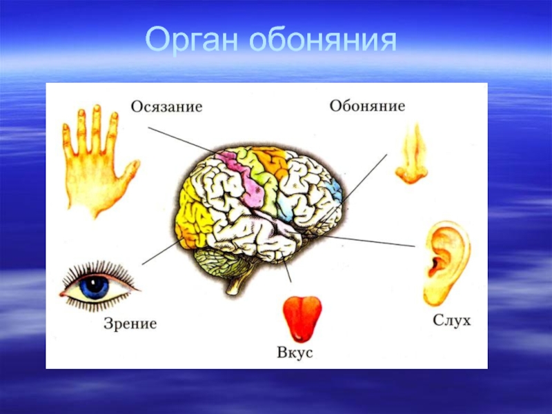

Слайд 2Differons are kept during all life and provide updating tissues. The

induction of a cell differentiation occurs as well - by means of contact with reacting system or on distance.
Under influence of external factors a regulation of duplication and a differentiation can be broken in a tissue, cells lose special properties and infinitely are made multiple copies, develops metaplasia of a tissue, then a tumour.

Слайд 3 Свойства тканей

Properties of tissues

Органная
специфичность
Organ specificity

Разная потребность
в кислороде,
питательных
веществах.
Different need in oxygen,
nutritious substances.

- Различная скорость
обмена веществ
Various speed of metabolism


Способность
к регенерации
Ability to
regeneration

Физиологическая
регенерация
(обновление).
Physiological regeneration
(renovaring).

Репаративная
регенерация
(восстановление
после повреждения)
Reparative regeneration
(restoration after damage)

Изменчивость
(пластичность)
Variability
(plasticity)

Адаптация к
изменившимся
условиям.
Adaptation to the
changed conditions.

Возрастные
изменения.
Age changes


Слайд 4Mesenchyme
It is an embryonic tissue from mobile cells with shoots and

jellylike intercellular substance.
Cells form little cell-islands - germs.
Germs are developed in different tissues: blood, a lymph, connecting tissues, a smooth muscular tissue depending on a microenvironment.

Слайд 5Эктодерма
Нервная
трубка
nervous tube
Хорда
chorda
Мезенхима
Mesenchyme
Мезенхима
Mesenchyme
Мезенхима
Mesenchyme
МЕЗЕНХИМА
(Mesenchyme)
стенки
желточного мешка
(yolk bag wall)


Слайд 6Ткани мезенхимного происхождения (опорно-трофические ткани)
развиваются из мезенхимы (are developed from mesenchyme)
состоят из

клеток и межклеточного вещества
(consist of cells and intercellular substance)
имеют единые стволовые клетки (have uniform stell cells )
отличаются большим разнообразием клеток (have the big variety of cells)
выполняют опорную, трофическую, защитную функции
(execute strong, trophic and protective functions)
обладают пластичностью и высокой способностью к регенерации и адаптации
(possess high ability to variability, regeneration and adaptation)

Tissues by mesenchyme origin


Слайд 7Система крови:
Органы, производящие компоненты крови (органы кроветворения и печень)
Кровь и лимфа

в сосудах, клетки крови в других тканях
3. Органы, в которых разрушаются компоненты крови (селезенка и печень)

System of blood:

Organs making blood
(haemopoietic organs
make blood cells, a liver –
proteins of plasma)

2. Peripheral blood and
a lymph, and also
blood cells in tissues

3. Organs destroy
components of blood
(a spleen and a liver).


Слайд 8Функции крови (Functions of blood) :
Трофическая (trophic)
Экскреторная (excretion)
Дыхательная (respiratory)
Защитная

(protective)
Гуморальная, регуляторная
(regulation over humor)
Гомеостатическая (homeostatic)

Кровь у взрослых – 6-8 % от массы тела,
(adult blood - 6-8 % from weight of a body)
у детей (children blood) – 9 %,
у новорожденных (newborns blood) – 13-15 %


Слайд 9Плазма крови (50-60%) Plasma of blood (50-60%)

90% воды и
10% сухого остатка

6,6-8,5% белки:
альбумины 4-4,5%
глобулины 2-3%
фибриноген
и протромбин 0,2-0,4%
белки комплемента

90% of water and
10% of the dry rest

6,6-8,5% proteins:
albumins 4-4,5%
globulins 2-3%
fibrinogen and
protrombin 0,2-0,4%
complement proteins


Слайд 10Erythrocyte
Thrombocyte (Platelet)
Leukocyte
Blood elements having form


Слайд 11Гемограмма (Hemogram) :

Эритроцитов (Erythrocytes) 4-5,5 x 10¹² / л
Лейкоцитов (Leukocytes)

3-10 x 109 / л
Тромбоцитов (Thrombocytes) 130-400 x 109/ л
Гематокрит (Hematocrit) , Ht - 30-35%
(доля форменных элементов) (part of form elements)
Гемоглобин (Hemoglobin) , Hb - 130-160 г/л
СОЭ (скорость оседания эритроцитов) 4-20 мм/час
SES (Speed of erythrocyte subsidence) (mm/hour)

Слайд 12Тромбоциты – фрагменты цитоплазмы мегакариоцитов
Thrombocytes (Platelets) - fragments of megakaryocyte’s

cytoplasm

Гиаломер
Hyalomere
(contains many
microtubules)

Хромомер
Granulomere
(granules containing
histamine,
serotonine,
thromboplastine,
enzymes,
regulatory proteins,
ions Са and Mg)

The size 2-3 µ, the form oval, can be shoots, live 10 days, die in a spleen and lungs.

At activation of blood coagulating factors thrombocytes stick together (agglutination)
and are once destructed, put out contents of granules and
start reactions of blood coagulation.


Слайд 13Эритроциты-дискоциты
Erythrocytes - diskocytes
a biconcave disk


Слайд 14Типы гемоглобина
Types of hemoglobin
Hb A

Hb F
(у взрослых 98%) (at adult 98%)
(фетальный,

у новорожденных 80%)
(fetal, at newborns 80%)

Hb + О2 = окси- Hb
oxi-Hb

В легких
into lungs

Ярко-красного
цвета
(артериальная кровь)
(arterial blood)

О2 + дезокси-Hb
desoxi-Hb

В тканях
into tissues

Вишневого
цвета
(венозная кровь)
(venous blood)

СО + Hb = карбокси-Hb (carboxi-Hb)

Угарный газ
carbonic oxide

Стойкое соединение
proof junction

При отравлении угарным газом (At a poisoning with carbonic oxide) :


Слайд 15Нормальный состав эритроцитов
Normal composition of blood erythrocytes
Ретикулоциты (reticulocytes)
Дискоциты (diskocytes) 80-90

%, нормоциты (normocytes) 75 %

Пойкилоциты (poikilocytes)10-20 %

0,2 – 1 %

Микроциты, макроциты
(microcytes, macrocytes) 25 %

Poikilocytosis is the increase
in number of the different form
erythrocytes.

Anisocytosis is the increase in number
of micro- and macro-erythrocytes.


Слайд 16Анизоцитоз и пойкилоцитоз в сканирующем электронном микроскопе
Anisocytosis and poikilocytosis at scanning

electron microskope

Нормоциты
Normocyte

Макроцит
Macrocyte

Эхиноцит
Echinocyte

Стоматоцит
Stomatocyte

Сфероцит
Spherocyte

Эхиноциты
Echinocytes


Слайд 17Пойкилоцитоз и анизоцитоз на мазке крови
Poikilocytosis and Anisocytosis at blood

smear

Пойкилоциты
poikilocytes

Микроцит
microcyte

Нормоцит
Normo-
cyte

Мегалоцит
megalocyte

Микроцит
microcyte

Макроцит
macrocyte


Слайд 18Количество эритроцитов
The quantity of erythrocytes :
(depends on need of an organism

in О2)

У мужчин (at man) – 3,9 – 5,5 х 1012 / литр

У женщин (at woman) – 3,5 – 5,0 х 1012 / литр

У детей до 1 года и в старости (at children to one year and old men) – 6 – 8 х 1012 / литр


Erythrocytes live 2-3 months, only in vessels, die in a spleen.
In hypotonic environment they swell and burst,
it is called Hemolisis.


Обратная связь

Если не удалось найти и скачать презентацию, Вы можете заказать его на нашем сайте. Мы постараемся найти нужный Вам материал и отправим по электронной почте. Не стесняйтесь обращаться к нам, если у вас возникли вопросы или пожелания:

Email: Нажмите что бы посмотреть 

Что такое ThePresentation.ru?

Это сайт презентаций, докладов, проектов, шаблонов в формате PowerPoint. Мы помогаем школьникам, студентам, учителям, преподавателям хранить и обмениваться учебными материалами с другими пользователями.


Для правообладателей

Яндекс.Метрика