- Главная
- Разное
- Дизайн
- Бизнес и предпринимательство
- Аналитика
- Образование
- Развлечения
- Красота и здоровье
- Финансы
- Государство
- Путешествия
- Спорт
- Недвижимость
- Армия
- Графика
- Культурология
- Еда и кулинария
- Лингвистика
- Английский язык
- Астрономия
- Алгебра
- Биология
- География
- Детские презентации
- Информатика
- История
- Литература
- Маркетинг
- Математика
- Медицина
- Менеджмент
- Музыка
- МХК
- Немецкий язык
- ОБЖ
- Обществознание
- Окружающий мир
- Педагогика
- Русский язык
- Технология
- Физика
- Философия
- Химия
- Шаблоны, картинки для презентаций
- Экология
- Экономика
- Юриспруденция
Генетична карта поліомавірусів dsDNA презентация
Содержание
- 1. Генетична карта поліомавірусів dsDNA
- 2. Генетична карта поліомавірусів вірусу SV-40 вірусу поліоми
- 3. Genome Organization of SV40 Early unit:
- 4. Functional organization of SV40 large T antigen
- 5. SV 40 DNA replication In the nucleus.
- 6. The replication of SV40 DNA
- 7. Genomic Organization of Papillomavirus Genome 7Kb circular
- 8. Early Region E2 (Regulator of DNA replication
- 9. Early Region Functional Assignments of Viral Early Proteins
- 10. Modulation of growth regulatory mechanisms Early Region Functional Assignments of Viral Early Proteins
- 15. Adenovirus Replication
- 16. Adenovirus Replication
- 17. Adenovirus Replication
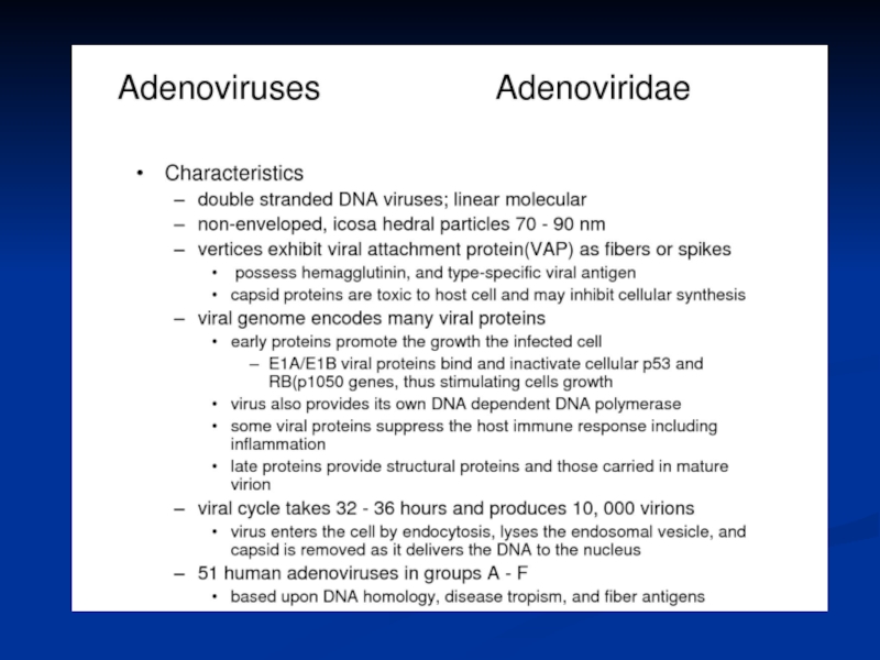
- 18. The early protein include: For transcription
- 19. Генетична карта вірусу простого герпесу
- 20. Транскрипція вірусу простого герпесу
- 21. Реплікація ДНК вірусу простого герпесу
- 22. Блочний запис генетичної інформації у герпесвірусів
- 23. Генетична карта бактеріофагу Т4
- 24. Схема розрізання конкатемеру фага Т4 Конкатемер ДНК
- 25. Генетична карта бактеріофагу лямбда
- 26. Інтеграція геному фагу лямбда
- 27. POXVIRIDAE Figure 2 Schematic representation of the
- 28. Figure 3 The infectious cycle
- 29. The poxvirus genome comprises a
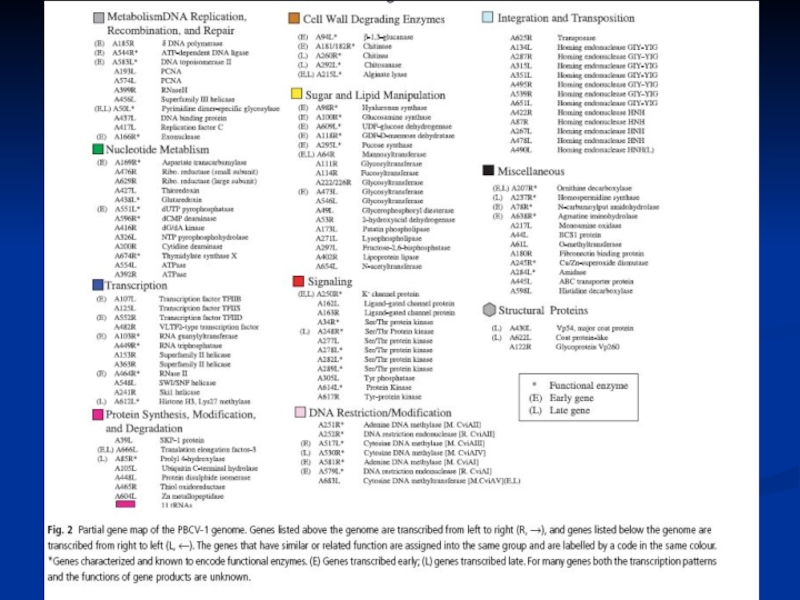
- 30. Генетична карта вірусу PBCV-1


Слайд 3Genome Organization of SV40
Early unit: large T antigen (LT), small t
antigen (sT), 17K T antigen (17KT).
Late unit: VP1, VP2, VP3 ,the agnoprotein (agno) and a pre-microRNA (miRNA).
Late unit: VP1, VP2, VP3 ,the agnoprotein (agno) and a pre-microRNA (miRNA).
The regulatory region (ori)
- For the early and late promoter and the origin of replication.
, small t antigen (sT), 17K T")
Слайд 4Functional organization of SV40 large T antigen
1. Activation of cellular DNA
and RNA synthesis
binding the cellular Rb and p53
causes the infected cell to begin a round of DNA replication.
2. Blockage of apoptosis
inactivate p53 at inappropiate times in the cell cycle.
3. Binding to the SV40 ori to initiate viral DNA replication
4. Shutting off early viral transcription
5. Activating late transcription
6. Virion assembly

Слайд 5SV 40 DNA replication
In the nucleus.
Large T antigen binds to
the SV40 origin
Using the host cell DNA polymerase,
which recognizes the viral origin of replication if the T antigen is present.
Bi-directional replication
Host histones complex with the newly made DNA.
Using the host cell DNA polymerase,
which recognizes the viral origin of replication if the T antigen is present.
Bi-directional replication
Host histones complex with the newly made DNA.


Слайд 7Genomic Organization of Papillomavirus
Genome
7Kb circular genome
Encode all proteins on the
same DNA strand
RNAs splicing
Long control region (LCR) contains transcriptional and replication regulatory elements
RNAs splicing
Long control region (LCR) contains transcriptional and replication regulatory elements

Слайд 8Early Region
E2 (Regulator of DNA replication and RNA transcription)
Functional Assignments of
Viral Early Proteins
Functional Assignments of Viral Early Proteins")

Слайд 10Modulation of growth regulatory mechanisms
Early Region
Functional Assignments of Viral Early Proteins

Слайд 15Adenovirus Replication
Endosome
Virion
Integrins
CAR
Virion DNA
Nuclear
pore
Nucleus
Cytoplasm
coxsackie-adenovirus receptor

Слайд 16Adenovirus Replication
Endosome
Early mRNAs
Early
proteins
Virion
Integrins
CAR
Virion DNA
Modulate Cell Cycle
Shut down host mRNA transport
Block CTL
Response
Nuclear
pore
Nucleus
Cytoplasm

Слайд 17Adenovirus Replication
Endosome
Late mRNAs
Early mRNAs
Early
proteins
Late
proteins
Virion
Integrins
CAR
Virion DNA
Modulate Cell Cycle
Shut down host mRNA transport
Progeny
DNA
Cell Lysis
Block CTL Response
Progeny
Virus
Nuclear
pore
Nucleus
Cytoplasm

Слайд 18The early protein include:
For transcription
EIA is referred to as an
‘immediate early’ gene)
For adenovirus DNA synthesis
viral DNA polymerase)
Alter expression of host cell genes
Against the host anti-viral response and/or interfere with cell cycle regulation
For adenovirus DNA synthesis
viral DNA polymerase)
Alter expression of host cell genes
Against the host anti-viral response and/or interfere with cell cycle regulation
For adenovirus")


Слайд 24Схема розрізання конкатемеру фага Т4
Конкатемер ДНК фагу Т4
Кінцеві надлишки
Розрізання
Геном фагу, що
пакується
в головку
в головку
Сайти розрізання
Мінімальний геном фагу
Фагова прегеномна ДНК являє собою багато копій геномної ДНК, з’єднаних “кінець-в-кінець”
Геном кожного фагу має циркулярні пермутації в лінійній генетичній карті
Геном кожного фагу має більше інформації, ніж мінімвльний геном


Слайд 27POXVIRIDAE
Figure 2 Schematic representation of the DNA of Vaccinia virus (VACV)
(WR strain): (Top) Linear double-stranded molecule with terminal hairpins and inverted repeats (not to scale). The denatured DNA forms a single-stranded circular molecule. (Bottom) Hind III cleavage sites of the Vaccinia virus (WR strain) genome, the asterisk indicates the fragment that contains the thymidine kinase gene used in construction of phylogenetic trees. Each 10-kbp terminal portion includes two groups of tandem repeats of short sequences rich in AT. (Redrawn from Fenner, Wittek, and Dumbell, 1989, with permission.)
 (WR strain): (Top) Linear")
Слайд 28
Figure 3 The infectious cycle of Vaccinia virus (VACV): IMV,
intracellular mature virus; EEV, extracellular enveloped virus; VITF, vaccinia intermediate transcription factor; VLTF, vaccinia late transcription factor; VETF, vaccinia early transcription factor.
: IMV, intracellular mature virus; EEV,")
Слайд 29
The poxvirus genome comprises a linear molecule of dsDNA with
covalently closed termini; terminal hairpins constitute two isomeric, imperfectly paired, “flip-flop” DNA forms consisting of inverted complementary sequences. Variably sized, tandem repeat sequence arrays may or may not be present near the ends (Fig. 2The poxvirus genome comprises a linear molecule of dsDNA with covalently closed termini; terminal hairpins constitute two isomeric, imperfectly paired, “flip-flop” DNA forms consisting of inverted complementary sequences. Variably sized, tandem repeat sequence arrays may or may not be present near the ends (Fig. 2). Replication takes place predominately if not exclusively within the cytoplasm (Fig. 3). Entry into cells of intracellular virus (IMV) and extracellular enveloped virus (EEV) is suggested to be via different pathways. After virion adsorption, IMV entry into the host cell is by fusion between the plasma membrane after which cores are released into the cytoplasm and uncoated further. EEV entry, unlike IMV, may necessitate fusion with endosomal membranes to release the core.
Polyadenylated, capped primary mRNA transcripts, representing about 50% of the genome, are initially synthesized from both DNA strands by enzymes within the core, including a virus-encoded multisubunit RNA polymerase; transcripts are extruded from the core for translation by host ribosomes. During synthesis of early proteins, host macromolecular synthesis is inhibited. Virus reproduction ensues in the host cell cytoplasm, producing basophilic (B-type) inclusions termed “viroplasms” or “virus factories”. The genome contains closely spaced ORFs, lacking introns, some of which may partially overlap preceded by virus-specific promoters that temporally regulate transcription of three classes of genes. One class, the early genes, are expressed from partially uncoated virions prior to DNA replication (these encode many non-structural proteins, including enzymes involved in replicating the genome and modifying DNA, RNA, and proteins designed to neutralize the host response). Early genes also encode intermediate transcription factors. Intermediate genes (which encode late transcription factors), are expressed during the period of DNA replication and are required for subsequent late gene transcription. Finally, late genes are expressed during the post-replicative phase (these mainly encode virion structural proteins but also early transcription factors). Despite a cytoplasmic site of replication, there is mounting evidence for the requirement of host nuclear proteins in post-replicative transcription. The mRNAs are capped, polyadenylated at the 3 termini, but not spliced. Many intermediate, late and some early mRNAs have 5 -poly(A) tracts, which precede the encoded mRNA. Early protein synthesis is generally decreased during the transition to late gene expression, but some genes are expressed from promoters with both early and late activity. Certain proteins are modified post-translationally (e.g., by proteolytic cleavage, phosphorylation, glycosylation, ribosylation, sulfation, acylation, palmitylation and myristylation). Proteolytic cleavage of late proteins is required for virion morphogenesis.
The replication of the DNA genome appears to be mainly through the action of viral enzymes. DNA replication appears to involve a self-priming, unidirectional, strand displacement mechanism in which concatemeric replicative intermediates are generated and subsequently resolved via specific cleavages into unit length DNAs that are ultimately covalently closed. Genetic recombination within genera has been shown, and may occur between daughter molecules during replication. Non-genetic genome reactivation generating infectious virus has been shown within and between genera of the Chordopoxvirinae.
Virus morphogenesis begins following DNA replication and expression of early, intermediate and late genes. Particle assembly is initiated with the formation of crescent-shaped membrane structures. Replicated, concatameric DNA is resolved into unit genomes and packaged into immature virion particles, which further mature to form intracellular mature virions (IMVs) which are fully infectious if liberated from cells. A portion of the IMVs are further enveloped by modified Golgi membranes, transported to the periphery of the cell where fusion of the wrapped virions with the plasma membrane ultimately results in release of extracellular enveloped virions (EEVs) by an as yet incompletely understood process. Enveloped virions thereby acquire host cell lipids and additional virus-specific proteins, including the virus hemagglutinin protein. The envelope is closely positioned to the surface membrane. While both IMVs and EEVs are infectious, the external antigens on the two virus forms are different and upon infection, the two forms of virus bind to different cellular receptors and are likely uncoated by different mechanisms. Virus DNA and several proteins are organized as a nucleoprotein complex within the core of all infectious virions. The IMVs contain an encompassing surface membrane, lateral bodies, and the nucleoprotein core complex (see Fig. 1). For Vaccinia virus, the core has a 9 nm thick membrane with a regular subunit structure. Within the vaccinia virion, negative stain indicates that the core assumes a biconcave shape (Fig. 1) apparently due to the large lateral bodies, although some evidence suggests the shape may represent an artifact of sample preparation. The lipoprotein surface membrane surrounding the Vaccinia virus core and lateral bodies is about 12 nm thick and contains irregularly shaped surface tubules composed of small globular subunits. During natural infections, the virus is likely spread by that population of virus released from the cells (EEV). Although the internal structure of Vaccinia virus is revealed in thin sections, the detailed internal structure of parapoxvirus particles is less evident (Fig. 1). In negatively stained preparations of parapoxviruses, superimposition of dorsal and ventral views of the surface filament sometimes produces a distinctive “criss-cross” surface appearance.