- Главная
- Разное
- Дизайн
- Бизнес и предпринимательство
- Аналитика
- Образование
- Развлечения
- Красота и здоровье
- Финансы
- Государство
- Путешествия
- Спорт
- Недвижимость
- Армия
- Графика
- Культурология
- Еда и кулинария
- Лингвистика
- Английский язык
- Астрономия
- Алгебра
- Биология
- География
- Детские презентации
- Информатика
- История
- Литература
- Маркетинг
- Математика
- Медицина
- Менеджмент
- Музыка
- МХК
- Немецкий язык
- ОБЖ
- Обществознание
- Окружающий мир
- Педагогика
- Русский язык
- Технология
- Физика
- Философия
- Химия
- Шаблоны, картинки для презентаций
- Экология
- Экономика
- Юриспруденция
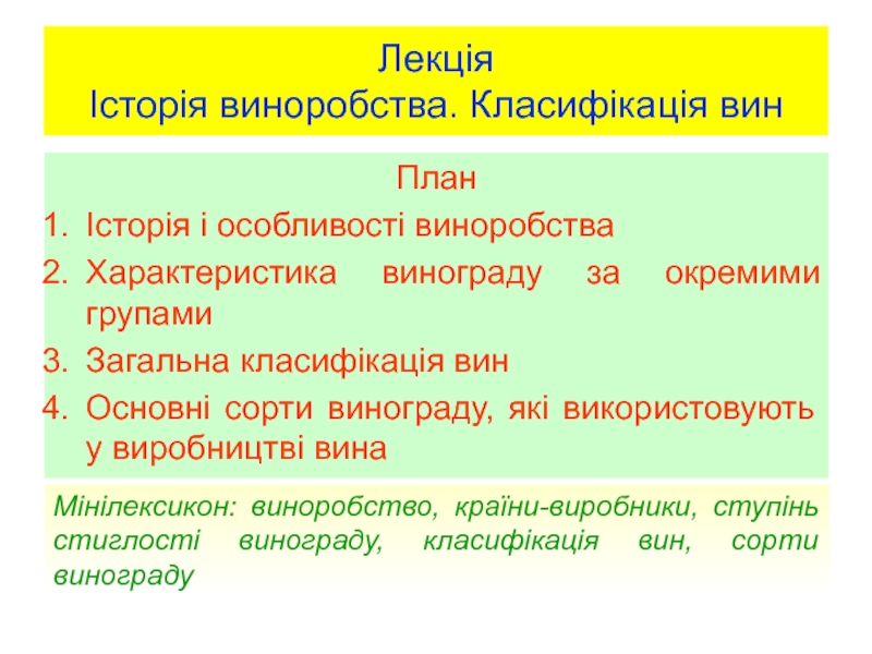
G-protein-coupled receptors презентация
Содержание
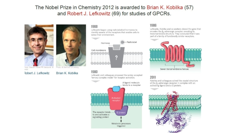
- 1. G-protein-coupled receptors
- 4. protein–coupled receptors are found only in
- 5. There are two principal signal transduction
- 6. Physiological roles GPCRs are involved in a
- 9. Structure - Single protein with 7 transmembrane regions
- 10. Ligands Monoamines e.g. dopamine,
- 11. Ligand binding site - varies depending
- 12. G-Protein alters shape GDP binding site distorted
- 13. Induced fit G-protein alters shape Complex
- 14. b) Interaction of αs with adenylate cyclase
- 17. Adrenoreceptor
- 18. Localization and the main effects α1- и
- 19. Механизм действия адренергических рецепторов. Эпинефрин и
- 21. Thank you for attention!!!

Слайд 4
protein–coupled receptors are found only in eukaryotes, including yeast, and animals.
The ligands that bind and activate these receptors include light-sensitive compounds, odors, pheromones, hormones, and neurotransmitters, and vary in size from small molecules to peptides to large proteins.
G protein–coupled receptors are involved in many diseases, and are also the target of approximately 40% of all modern medicinal drugs.

Слайд 5
There are two principal signal transduction pathways involving the G protein–coupled
the cAMP signal pathway and
the phosphatidylinositol signal pathway

Слайд 6Physiological roles GPCRs are involved in a wide variety of physiological processes.
The visual sense: The opsins use a photoisomerization reaction to translate electromagnetic radiation into cellular signals. Rhodopsin, for example, uses the conversion of 11-cis-retinal to all-trans-retinal for this purpose
The gustatory sense (taste): GPCRs in taste cells mediate release of gustducin in response to bitter- and sweet-tasting substances.
The sense of smell: Receptors of the olfactory epithelium bind odorants (olfactory receptors) and pheromones (vomeronasal receptors)
Behavioral and mood regulation: Receptors in the mammalian brain bind several different neurotransmitters, including serotonin, dopamine, GABA, and glutamate
Regulation of immune system activity and inflammation: Chemokine receptors bind ligands that mediate intercellular communication between cells of the immune system; receptors such as histamine receptors bind inflammatory mediators and engage target cell types in the inflammatory response. GPCRs are also involved in immune-modulation and directly involved in suppression of TLR-induced immune responses from T cells.
Autonomic nervous system transmission: Both the sympathetic and parasympathetic nervous systems are regulated by GPCR pathways, responsible for control of many automatic functions of the body such as blood pressure, heart rate, and digestive processes
Cell density sensing: A novel GPCR role in regulating cell density sensing.
Homeostasis modulation (e.g., water balance).
Involved in growth and metastasis of some types of tumors.


Слайд 10 Ligands
Monoamines e.g. dopamine, histamine, noradrenaline, acetylcholine (muscarinic)
Nucleotides
Lipids
Hormones
Glutamate
Ca++
G-protein-coupled
NucleotidesLipidsHormonesGlutamateCa++ G-protein-coupled receptors (7-TM receptors)")
Слайд 11 Ligand binding site - varies depending on receptor type
A) Monoamines
B) Peptide hormones - top of TM helices + extracellular loops + N-terminal chain
C) Hormones - extracellular loops + N-terminal chain
D) Glutamate - N-terminal chain
G-protein-coupled receptors (7-TM receptors)
 Monoamines - pocket in TM")
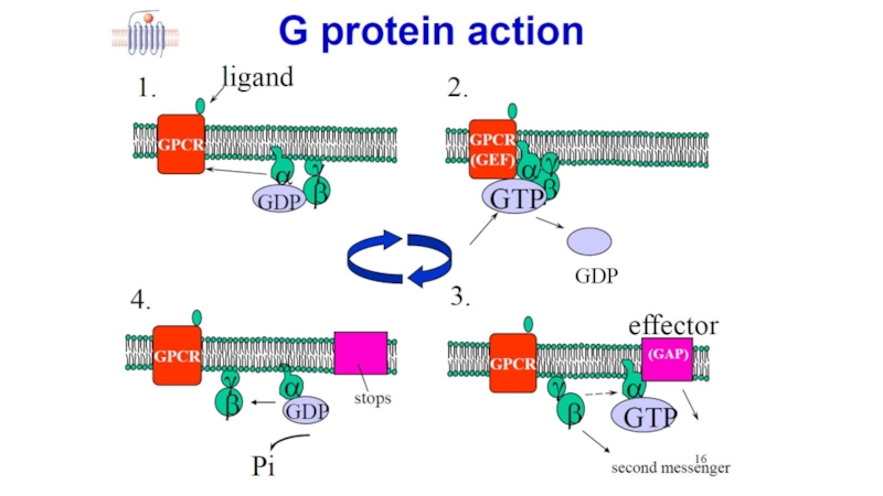
Слайд 12G-Protein alters shape
GDP binding site distorted
GDP binding weakened
GDP departs
a) Interaction of
3. G-protein-coupled receptors (7-TM receptors)
3.6 Signal transduction pathway
 Interaction of receptor with Gs-protein3. G-protein-coupled")
Слайд 13Induced fit
G-protein alters shape
Complex destabilised
Process repeated for as long
Signal amplification - several G-proteins activated by one ligand
αs Subunit carries message to next stage
a) Interaction of receptor with Gs-protein
3. G-protein-coupled receptors (7-TM receptors)
3.6 Signal transduction pathway

Слайд 14b) Interaction of αs with adenylate cyclase
3. G-protein-coupled receptors (7-TM
3.6 Signal transduction pathway
 Interaction of αs with adenylate cyclase3. G-protein-coupled receptors (7-TM receptors)3.6 Signal transduction pathway")

Слайд 18Localization and the main effects
α1- и β1- receptors localized mainly in
α2- и β2- receptors are extrasynaptic, and are also available on the presynaptic membrane of the same neurons. On the α2-receptors act as adrenaline and noradrenaline. β2-receptors are sensitive mainly to adrenaline. Α2-receptors on the presynaptic membrane noradrenaline acts on the principle of negative feedback - inhibits proper selection .
α1 — localized in arterioles, stimulation leads to a spasm of arterioles, increasing the pressure, decrease vascular permeability and a decrease in exudative inflammation.
α2 — mainly presynaptic receptors are "negative feedback loop" for the adrenergic system and their stimulation leads to lower blood pressure
β1 — localized in the heart, the stimulation frequency leads to an increase (positive chronotropic effect) and force of cardiac contractions (positive inotropic effect) in addition, increases the myocardial oxygen consumption and increase blood pressure. It is also localized in the kidneys, being receptors juxtaglomerular apparatus.
β2 — located in the bronchioles, the stimulation causes dilation of the bronchial tubes and the removal of bronchospasm. These receptors are found on cells of the liver, the effects on them hormone causes glycogenolysis and glucose output in blood.
β3 — located in the adipose tissue. Stimulation of these receptors enhances lipolysis and leads to the release of energy and to increase heat production

Слайд 19
Механизм действия адренергических рецепторов. Эпинефрин и норадреналин являются лигандами для адренергических