- Главная
- Разное
- Дизайн
- Бизнес и предпринимательство
- Аналитика
- Образование
- Развлечения
- Красота и здоровье
- Финансы
- Государство
- Путешествия
- Спорт
- Недвижимость
- Армия
- Графика
- Культурология
- Еда и кулинария
- Лингвистика
- Английский язык
- Астрономия
- Алгебра
- Биология
- География
- Детские презентации
- Информатика
- История
- Литература
- Маркетинг
- Математика
- Медицина
- Менеджмент
- Музыка
- МХК
- Немецкий язык
- ОБЖ
- Обществознание
- Окружающий мир
- Педагогика
- Русский язык
- Технология
- Физика
- Философия
- Химия
- Шаблоны, картинки для презентаций
- Экология
- Экономика
- Юриспруденция
Fertilization and development fertilization презентация
Содержание
- 1. Fertilization and development fertilization
- 2. Copyright Pearson Prentice Hall What is fertilization? Fertilization
- 3. Copyright Pearson Prentice Hall Fertilization The
- 4. Copyright Pearson Prentice Hall Fertilization After the
- 5. Copyright Pearson Prentice Hall Early Development Early
- 6. Copyright Pearson Prentice Hall Early Development What are the stages of early development?
- 7. Copyright Pearson Prentice Hall Early Development
- 8. Copyright Pearson Prentice Hall Early Development Implantation
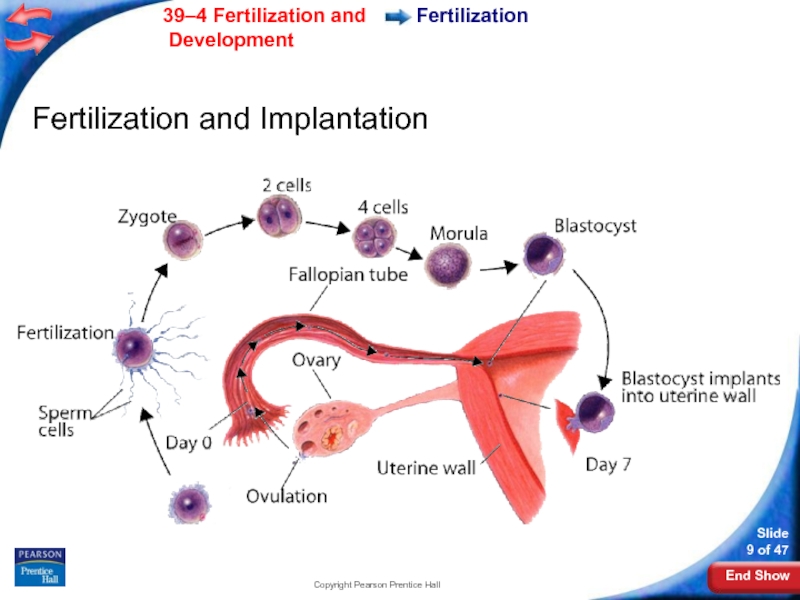
- 9. Copyright Pearson Prentice Hall Fertilization Fertilization and Implantation
- 10. Copyright Pearson Prentice Hall Early Development Blastocyst
- 11. Copyright Pearson Prentice Hall Early Development A
- 12. Copyright Pearson Prentice Hall Early Development Gastrulation
- 13. Copyright Pearson Prentice Hall Mesoderm Amniotic cavity
- 14. Copyright Pearson Prentice Hall Early Development The
- 15. Copyright Pearson Prentice Hall Early Development The
- 16. Copyright Pearson Prentice Hall Early Development Neurulation
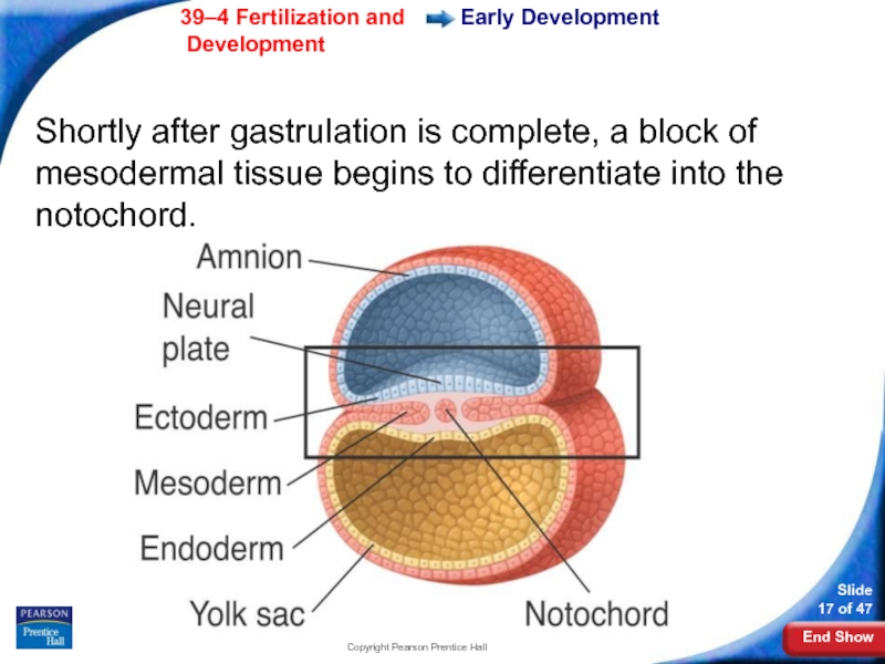
- 17. Copyright Pearson Prentice Hall Early Development Shortly
- 18. Copyright Pearson Prentice Hall Neural crest Neural
- 19. Copyright Pearson Prentice Hall Neural crest Neural
- 20. Copyright Pearson Prentice Hall Early Development Extraembryonic
- 21. Copyright Pearson Prentice Hall Early Development The
- 22. Copyright Pearson Prentice Hall Fingerlike projections called
- 23. Copyright Pearson Prentice Hall The chorionic villi
- 24. Copyright Pearson Prentice Hall Early Development What is the function of the placenta?
- 25. Copyright Pearson Prentice Hall Early Development
- 26. Copyright Pearson Prentice Hall Early Development The
- 27. Copyright Pearson Prentice Hall Early Development After
- 28. Copyright Pearson Prentice Hall Control of Development
- 29. Copyright Pearson Prentice Hall Later Development Later
- 30. Copyright Pearson Prentice Hall Later Development During
- 31. Copyright Pearson Prentice Hall Childbirth Childbirth About
- 32. Copyright Pearson Prentice Hall Childbirth The mother’s
- 33. Copyright Pearson Prentice Hall Childbirth The opening
- 34. Copyright Pearson Prentice Hall Childbirth The baby
- 35. Copyright Pearson Prentice Hall Multiple Births Multiple
- 36. Copyright Pearson Prentice Hall Early Years Early
- 37. Copyright Pearson Prentice Hall Adulthood Adulthood Development
- 38. Copyright Pearson Prentice Hall 39–4
- 39. Copyright Pearson Prentice Hall 39–4 Fertilization takes
- 40. Copyright Pearson Prentice Hall 39–4 The process
- 41. Copyright Pearson Prentice Hall 39–4 The central
- 42. Copyright Pearson Prentice Hall 39–4 The placenta
- 43. Copyright Pearson Prentice Hall 39–4 Which of
- 44. END OF SECTION
Слайд 3Copyright Pearson Prentice Hall
Fertilization
The process of a sperm joining an egg
Слайд 4Copyright Pearson Prentice Hall
Fertilization
After the two haploid (N) nuclei fuse, a
A diploid cell has a set of chromosomes from each parent cell.
The fertilized egg is called a zygote.
Слайд 5Copyright Pearson Prentice Hall
Early Development
Early Development
While still in the Fallopian tube,
Four days after fertilization, the embryo is a solid ball of about 64 cells called a morula.
Слайд 7Copyright Pearson Prentice Hall
Early Development
The stages of early development include implantation,
Слайд 8Copyright Pearson Prentice Hall
Early Development
Implantation
As the morula grows, it becomes a
6–7 days after fertilization, the blastocyst attaches to the uterine wall.
The embryo secretes enzymes that digest a path into it.
This process is known as implantation.
Слайд 10Copyright Pearson Prentice Hall
Early Development
Blastocyst cells specialize due to the activation
This process, called differentiation, is responsible for the development of the various types of tissue in the body.
Слайд 11Copyright Pearson Prentice Hall
Early Development
A cluster of cells, known as the
The embryo will develop from these cells, while the other cells will differentiate into tissues that surround the embryo.
Слайд 12Copyright Pearson Prentice Hall
Early Development
Gastrulation
The inner cell mass of the blastocyst
Слайд 13Copyright Pearson Prentice Hall
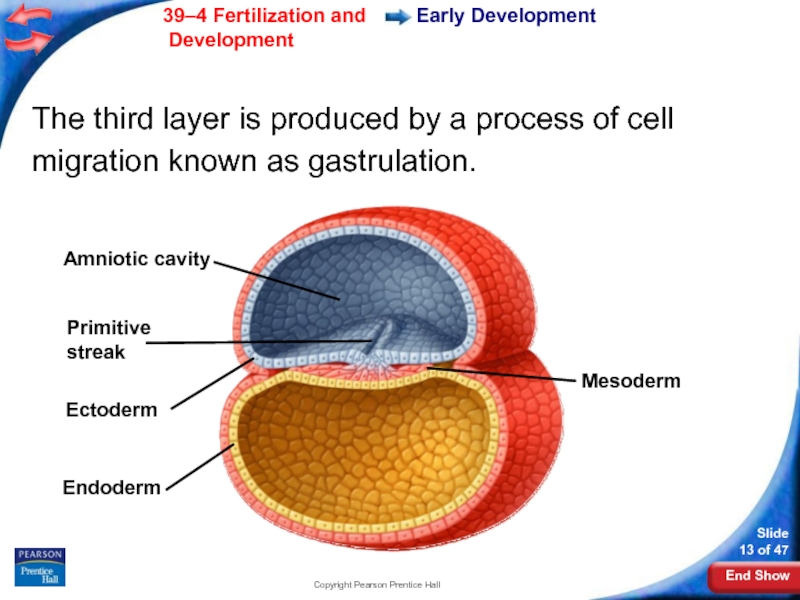
Mesoderm
Amniotic cavity
Primitive streak
Ectoderm
Endoderm
Early Development
The third layer is produced
Слайд 14Copyright Pearson Prentice Hall
Early Development
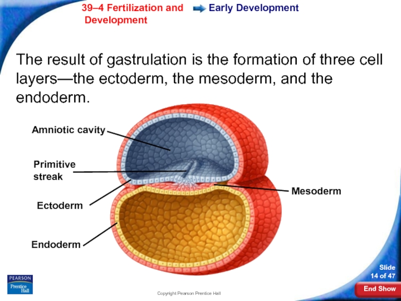
The result of gastrulation is the formation
Amniotic cavity
Primitive streak
Ectoderm
Endoderm
Mesoderm
Слайд 15Copyright Pearson Prentice Hall
Early Development
The ectoderm develops into the skin and
The endoderm forms the digestive lining and organs.
Mesoderm cells differentiate into internal tissues and organs.
Слайд 16Copyright Pearson Prentice Hall
Early Development
Neurulation
Gastrulation is followed by neurulation.
Neurulation is
Слайд 17Copyright Pearson Prentice Hall
Early Development
Shortly after gastrulation is complete, a block
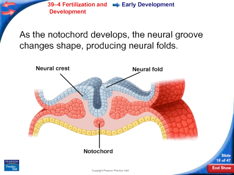
Слайд 18Copyright Pearson Prentice Hall
Neural crest
Neural fold
Notochord
Early Development
As the notochord develops, the
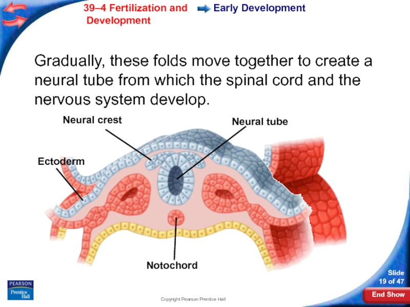
Слайд 19Copyright Pearson Prentice Hall
Neural crest
Neural tube
Ectoderm
Notochord
Early Development
Gradually, these folds move together
Слайд 20Copyright Pearson Prentice Hall
Early Development
Extraembryonic Membranes
As the embryo develops, membranes form
Two of these membranes are the amnion and the chorion.
Слайд 21Copyright Pearson Prentice Hall
Early Development
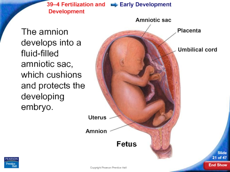
The amnion develops into a fluid-filled amniotic
Uterus
Amnion
Fetus
Amniotic sac
Placenta
Umbilical cord
Слайд 22Copyright Pearson Prentice Hall
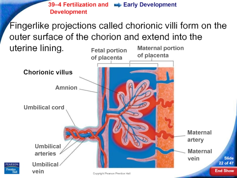
Fingerlike projections called chorionic villi form on the
Early Development
Fetal portion of placenta
Maternal portion of placenta
Maternal artery
Maternal vein
Umbilical vein
Umbilical arteries
Umbilical cord
Amnion
Chorionic villus
Слайд 23Copyright Pearson Prentice Hall
The chorionic villi and uterine lining form the
The placenta connects the mother and developing embryo.
Early Development
Слайд 25Copyright Pearson Prentice Hall
Early Development
The placenta is the embryo's organ of
Слайд 26Copyright Pearson Prentice Hall
Early Development
The placenta acts as a barrier to
Some disease causing agents, such as German measles and HIV can cross the placenta.
Some drugs, including alcohol and medications also can penetrate the placenta and affect development.
Слайд 27Copyright Pearson Prentice Hall
Early Development
After eight weeks, the embryo is called
After three months, most major organs and tissues are formed. During this time, the umbilical cord also forms.
The umbilical cord connects the fetus to the placenta.
Слайд 28Copyright Pearson Prentice Hall
Control of Development
Control of Development
The fates of many
The inner cell mass contains embryonic stem cells, unspecialized cells that can differentiate into nearly any specialized cell type.
Researchers are still learning the mechanisms that control stem cell differentiation.
Слайд 29Copyright Pearson Prentice Hall
Later Development
Later Development
4–6 months after fertilization:
The heart can
Bone replaces cartilage that forms the early skeleton.
A layer of soft hair grows over the fetus’s skin.
The fetus grows and the mother can feel it moving.
Слайд 30Copyright Pearson Prentice Hall
Later Development
During the last three months, the organ
The fetus doubles in mass.
It can now regulate its body temperature.
The central nervous system and lungs completely develop.
Слайд 31Copyright Pearson Prentice Hall
Childbirth
Childbirth
About nine months after fertilization, the fetus is
A complex set of factors affects the onset of childbirth.
Слайд 32Copyright Pearson Prentice Hall
Childbirth
The mother’s posterior pituitary gland releases the hormone
These muscles begin rhythmic contractions known as labor.
The contractions become more frequent and more powerful.
Слайд 33Copyright Pearson Prentice Hall
Childbirth
The opening of the cervix expands until it
At some point, the amniotic sac breaks, and the fluid it contains rushes out of the vagina.
Contractions force the baby out through the vagina.
Слайд 34Copyright Pearson Prentice Hall
Childbirth
The baby now begins an independent existence.
Its
Слайд 35Copyright Pearson Prentice Hall
Multiple Births
Multiple Births
If two eggs are released during
A single zygote may split apart to produce two embryos, which are called identical twins.
Слайд 36Copyright Pearson Prentice Hall
Early Years
Early Years
The first two years of life
Childhood lasts from infancy until puberty.
Adolescence begins with puberty and ends with adulthood.
Puberty produces a growth spurt that will conclude in mid-adolescence.
Слайд 37Copyright Pearson Prentice Hall
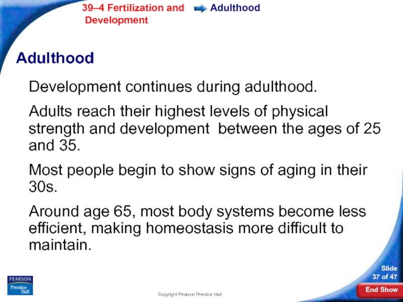
Adulthood
Adulthood
Development continues during adulthood.
Adults reach their highest
Most people begin to show signs of aging in their 30s.
Around age 65, most body systems become less efficient, making homeostasis more difficult to maintain.
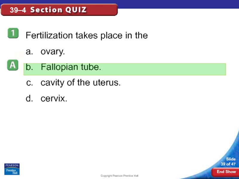
Слайд 39Copyright Pearson Prentice Hall
39–4
Fertilization takes place in the
ovary.
Fallopian tube.
cavity of the
cervix.
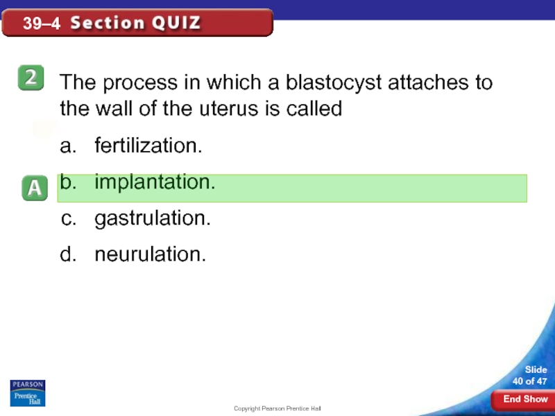
Слайд 40Copyright Pearson Prentice Hall
39–4
The process in which a blastocyst attaches to
fertilization.
implantation.
gastrulation.
neurulation.
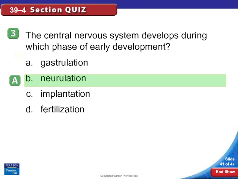
Слайд 41Copyright Pearson Prentice Hall
39–4
The central nervous system develops during which phase
gastrulation
neurulation
implantation
fertilization
Слайд 42Copyright Pearson Prentice Hall
39–4
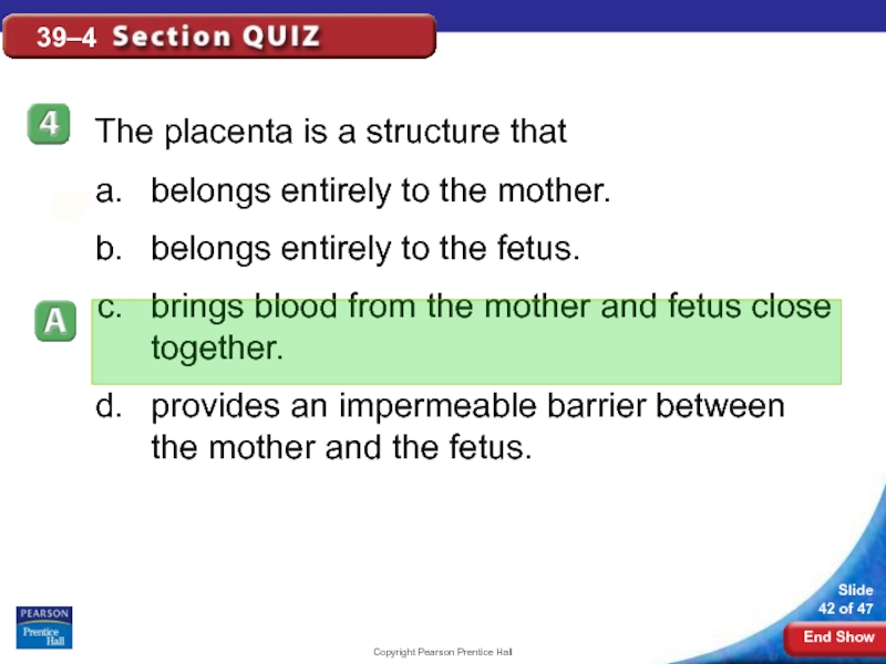
The placenta is a structure that
belongs entirely to
belongs entirely to the fetus.
brings blood from the mother and fetus close together.
provides an impermeable barrier between the mother and the fetus.
Слайд 43Copyright Pearson Prentice Hall
39–4
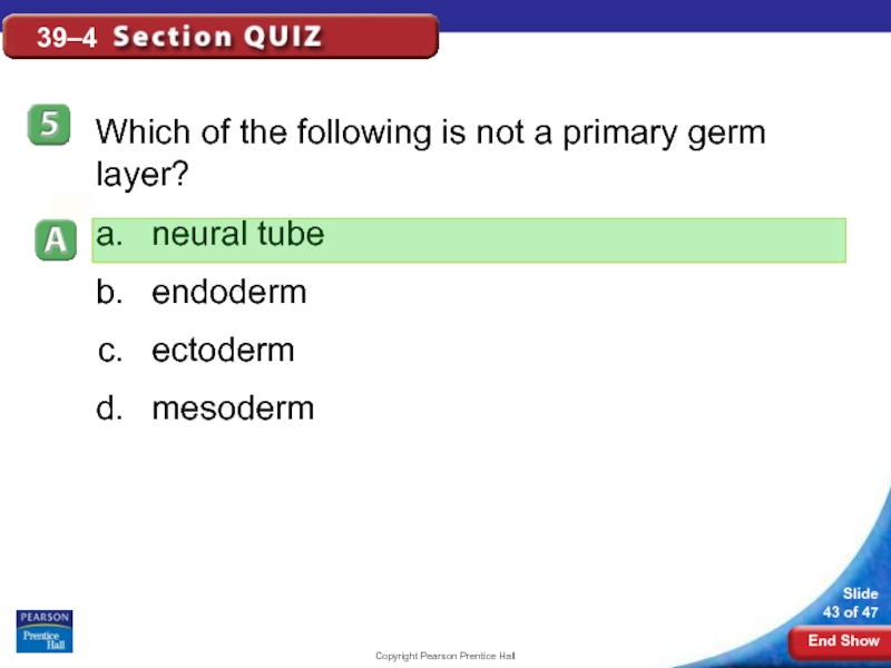
Which of the following is not a primary
neural tube
endoderm
ectoderm
mesoderm
