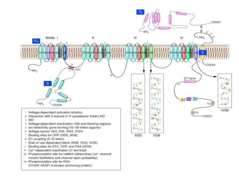


L-type Ca2+ channel. Shown is the pore-forming 1C subunit consisting of 4 homologous repeated domains (I–IV), each composed of 6 transmembrane segments as described in text. The cytoplasmic ß subunit is formed by 2 highly conserved domains indicated in purple, and the amino-terminal portion of the second conserved domain interacts with the I–II loop of 1C. The subunit has a single transmembrane segment with a short cytoplasmic C terminus and is linked by a disulfide bound to the extracellular, glycosylated 2 subunit. PKA phosphorylation sites of proven functional significance are shown as green diamonds at Ser1928 on 1C and Ser478 and Ser479 on ß2a. PKC phosphorylation sites of proven functional importance at Thr27 and Thr31 on 1C are indicated by yellow squares.
- Главная
- Разное
- Дизайн
- Бизнес и предпринимательство
- Аналитика
- Образование
- Развлечения
- Красота и здоровье
- Финансы
- Государство
- Путешествия
- Спорт
- Недвижимость
- Армия
- Графика
- Культурология
- Еда и кулинария
- Лингвистика
- Английский язык
- Астрономия
- Алгебра
- Биология
- География
- Детские презентации
- Информатика
- История
- Литература
- Маркетинг
- Математика
- Медицина
- Менеджмент
- Музыка
- МХК
- Немецкий язык
- ОБЖ
- Обществознание
- Окружающий мир
- Педагогика
- Русский язык
- Технология
- Физика
- Философия
- Химия
- Шаблоны, картинки для презентаций
- Экология
- Экономика
- Юриспруденция
Elektrofiziologiy презентация
Содержание

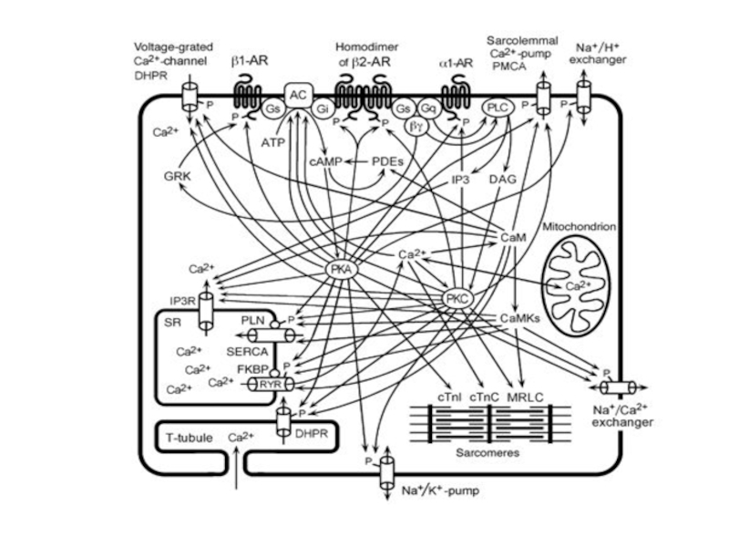
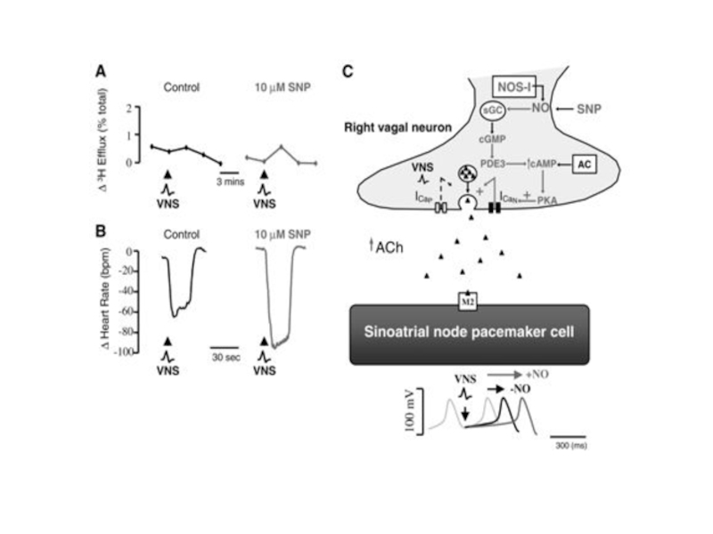
Слайд 45Signaling cascades regulating L-type Ca2+ channels. A, Schematic of the cAMP/PKA
cascade regulating L-type channels. Stimulation of ß1AR or ß2AR leads to Gs-mediated activation of AC and increased production of cAMP, which stimulates PKA, as described in text. PKA can then phosphorylate the channel at multiple potential sites indicated schematically by the single P in the diagram. The PKA phosphorylated site(s) is then sensitive to the phosphatases PP1 and PP2A. Whereas ß1AR regulation causes more global increases in cAMP, ß2AR stimulation can result in highly localized cAMP level changes and regulation. Regulatory proteins may be localized to the channel by an AKAP for PKA and by binding of PP2A to the C terminus of the channel. Muscarinic M2 receptors can oppose the ßAR upregulation of ICa by acting through Gi to inhibit AC. B, PLC/PKC signaling cascade regulating L-type Ca2+ channels. Activation of 1-adrenergic, ET, or AT1 receptors stimulates Gq with resulting activation of PLC, which leads to the production of diacylglycerol and activation of PKC. PKC is proposed to target to the membrane by binding a RACK protein in the vicinity of the L-type Ca2+ channel, which it then phosphorylates (see text for details). A Ser/Thr phosphatase counterbalances this phosphorylation. IP3 indicates inositol trisphosphate; PIP2, phosphatidylinositol 4,5 bisphosphate.