- Главная
- Разное
- Дизайн
- Бизнес и предпринимательство
- Аналитика
- Образование
- Развлечения
- Красота и здоровье
- Финансы
- Государство
- Путешествия
- Спорт
- Недвижимость
- Армия
- Графика
- Культурология
- Еда и кулинария
- Лингвистика
- Английский язык
- Астрономия
- Алгебра
- Биология
- География
- Детские презентации
- Информатика
- История
- Литература
- Маркетинг
- Математика
- Медицина
- Менеджмент
- Музыка
- МХК
- Немецкий язык
- ОБЖ
- Обществознание
- Окружающий мир
- Педагогика
- Русский язык
- Технология
- Физика
- Философия
- Химия
- Шаблоны, картинки для презентаций
- Экология
- Экономика
- Юриспруденция
Crispr Cas9 Genome Editing презентация
Содержание
- 1. Crispr Cas9 Genome Editing
- 2. CRISPR Gene-Editing Tool against Tumors Mesothelin-specific
- 3. The cells modified and express Chimeric
- 4. a, TRAC locus with the 5′ end (grey) of
- 5. CAR T cells created with CRISPR were
- 7. (a) Model of CRISPR/Cas9 directed intracellular defense
- 8. Figure 2 | CRISPR/Cas9 directed disruption of
- 9. In summary, these results indicate that
- 10. Crispr/Cas9-Based Genome editing for correction of Dystrophin Mutations that cause Duchenne Muscular Dystrophy
- 11. CRISPR/Cas9 targeting of the dystrophin gene
- 12. Gene editing capabilities of CRISPR/Cas9
- 13. References Eyquem, J., Mansilla-Soto, J., Giavridis,

Слайд 2CRISPR Gene-Editing Tool against Tumors
Mesothelin-specific CAR T cells attacking a cancer
cell.
Credit: Prasad Adusumilli / Memorial Sloan Kettering
Credit: Prasad Adusumilli / Memorial Sloan Kettering

Слайд 3
The cells modified and express Chimeric Antigen Receptors (CARs) on the
surface so that they can recognize and attact cancer cells.
CAR gene in the T-cell receptor alpha chain (TRAC) gene which includes the gene for the T-cell receptor
Effective at destroying tumor cells than those in which it was inserted randomly with a retrovirus
CAR gene in the T-cell receptor alpha chain (TRAC) gene which includes the gene for the T-cell receptor
Effective at destroying tumor cells than those in which it was inserted randomly with a retrovirus
 on the surface so that they")
Слайд 4a, TRAC locus with the 5′ end (grey) of the TRAC first exon, the TRAC gRNA (blue)
and the corresponding PAM sequence (red). The two blue arrows indicate the predicted Cas9 double strand break. Bottom, CRISPR/Cas9-targeted integration into the TRAC locus. The targeting construct (AAV) contains a splice acceptor (SA), followed by a P2A coding sequence, the 1928z CAR gene and a polyA sequence, flanked by sequences homologous to the TRAC locus (LHA and RHA, left and right homology arm). Once integrated, the endogenous TCRα promoter drives CAR expression, while the TRAC locus is disrupted. TRAV, TCRα variable region; TRAJ, TCRα joining region; 2A, the self-cleaving Porcine teschovirus 2A sequence. pA: bovine growth hormone polyA sequence.
b, Timeline of the CAR targeting into primary T cells.
c, Representative TCR/CAR flow plots 4 days after transfection of T cells with Cas9 mRNA and TRAC gRNA and addition of AAV6 at the indicated multiplicity of infection.
b, Timeline of the CAR targeting into primary T cells.
c, Representative TCR/CAR flow plots 4 days after transfection of T cells with Cas9 mRNA and TRAC gRNA and addition of AAV6 at the indicated multiplicity of infection.
 of the TRAC first exon, the TRAC gRNA (blue) and the corresponding PAM")
Слайд 5CAR T cells created with CRISPR were less likely to stop
recognizing and attacking tumor cells
prove safer than random integration
need not come from a patient's own T cells
easier and cheaper manufacture of CAR T cells.
implications for research on diseases other than cancer
prove safer than random integration
need not come from a patient's own T cells
easier and cheaper manufacture of CAR T cells.
implications for research on diseases other than cancer

Слайд 7(a) Model of CRISPR/Cas9 directed intracellular defense against lentiviral infection. hCas9
and its gRNA can be synthesized from either transfected plasmids (module A) or knockin expression cassettes (module B). After synthesis, hCas9 and gRNA assemble and work as an antiviral module in different steps. Once penetrating into the host cells, virally encoded reverse transcriptase uses the lentiviral RNA genome as a template for the synthesis of viral cDNA. Viral integrase binds and processes the LTR and assists in the insertion of the viral cDNA into the host genome. During these processes, the viral genome is unprotected by its envelope and capsid, rendering the viral cDNA vulnerable to cleavage by DNA endonucleases. In our study, the CRISPR/Cas9 system was adapted for human cells as an antiviral module, which bound and disrupted the viral genome of both pre-integration viral DNA (Step 1) and integrated provirus (Step 2).
(b) Seven gRNAs were designed and used with hCas9 to
target corresponding regions in the EGFP reporter lentivirus, including gEGFP-T1 to -T4 and gLTR-T1 to -T3. (c) FACS analysis of HEK293 cells that were pretreated with hCas9-mCh and gRNAs for 20B24 h, and then challenged by EGFP reporter lentiviruses for 4 days. gEmpty is an empty gRNA vector and gMock (as the gS35 in Supplementary Fig. 1) is a non-targeting mock gRNA.
(d–g) The CRISPR/Cas9 system can direct targeted disruption of the nonintegrative lentivirus.
(d) Fluorescence microscopy images of HEK293 cells pretreated with CRISPR/Cas9 followed by non-integrative lentivirus infection for 3 days. Insets show bright field images. Scale bar, 500 mm.
 Model of CRISPR/Cas9 directed intracellular defense against lentiviral infection. hCas9 and its gRNA can")
Слайд 8Figure 2 | CRISPR/Cas9 directed disruption of integrated lentivirus.CRISPR/Cas9-mediated disruption of
integrated lenti-proviral DNA in infected HEK293 cells.
(a) Top: confocal images of cells with high copy numbers of EGFP proviral DNA that were co-transfected with hCas9 and either gEGFP-T1 (right panels) or mock gRNA (middle panels). Left panels show untreated cells. Images were taken at 14 days after transfection. Bottom: confocal images with DAPI staining (blue). Scale bars, 50 mm
(b) Determination of provirus copy numbers in three different lentiviral integrated HEK293 cell lines.
(c) PCR analysis of proviral DNA. The amount of full-length provirus (arrow) was reduced in the GFP-negative cells generated by CRISPR/Cas9-mediated lentiviral disruption.
(d) DNA sequencing analysis of the CRISPR/Cas9 target sites in the EGFP proviruses. Two examples shown here were treated with hCas9 and either gEGFP-T1 (top) or gLTR-T2 (bottom). Sequences identified multiple times were marked accordingly on the right.
(e) Proviral copy number quantitation by qPCR. Statistical analysis determined using unpaired t-test (***Po0.001; **Po0.01; *Po0.1).
(f) FACS analysis of proviral EGFP expression
at different days after transfection of hCas9 and gRNAs into cells with either high or low provirus copy numbers as determined in b.

Слайд 9
In summary, these results indicate that the CRISPR/Cas9 system can mediate
targeted disruption of both pre-integration viruses and integrated proviruses with dsDNA in either linear or circular format.

Слайд 10Crispr/Cas9-Based Genome editing for correction of Dystrophin Mutations that cause Duchenne
Muscular Dystrophy

Слайд 11CRISPR/Cas9 targeting of the dystrophin gene (A) sgRNA sequences were designed
to bind sequences in the exon 45–55 mutational hotspot region of the dystrophin gene, such that gene editing could restore dystrophin expression from a wide variety of patient-specific mutations. Arrows within introns indicate sgRNA targets designed to delete entire exons from the genome. Arrows within exons indicate sgRNA targets designed to create targeted frameshifts in the dystrophin gene.
(B) Example of frame correction following introduction of small insertions or deletions by NHEJ DNA repair in exon 51 using the CR3 sgRNA.
(C) Schematic of multiplex sgRNA targets designed to delete exon 51 and restore the dystrophin reading frame in a patient mutation with the deletion of exons 48–50. (D) Schematic of multiplex sgRNA targets designed to delete the entire exon 45–55 region to address a variety of DMD patient mutations.
 sgRNA sequences were designed to bind sequences in")
Слайд 12
Gene editing capabilities of CRISPR/Cas9 system can correct up to 62%
of Duchenne Muscular Dystrophy
Collectively,this study provides proof-of-principle that the CRISPR/Cas9 technology is a versatile method for correcting a significant fraction of dystrophin mutations and with continued development may serve as a general platform for treating genetic disease.
Collectively,this study provides proof-of-principle that the CRISPR/Cas9 technology is a versatile method for correcting a significant fraction of dystrophin mutations and with continued development may serve as a general platform for treating genetic disease.

Слайд 13References
Eyquem, J., Mansilla-Soto, J., Giavridis, T., van der Stegen, S. J.
C., Hamieh, M., Cunanan, K. M., … Sadelain, M. (2017). Targeting a CAR to the TRAClocus with CRISPR/Cas9 enhances tumour rejection. Nature, 543(7643), 113–117. doi: 10.1038/nature21405.
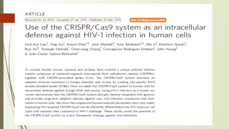
Liao, H. K., Gu, Y., Diaz A., Marlett, J., Takahashi, Y., Li, M.,… Sadelain, M. (2015). Use of the CRISPR/Cas9 system as an intracellular defense against HIV-1 infection in human cells . Nature Communications, 6:6413, 1–7. doi: 10.1038/ncomms7413.
David G. Ousterout, Ami M. Kabadi, Pratiksha I. Thakore, William H. Majoros, Timothy E. Reddy, and Charles A. Gersbach . Multiplex CRISPR/Cas9-Based Genome Editing for Correction of Dystrophin Mutations that Cause Duchenne Muscular Dystrophy/ Published in final edited form as: Nat Commun. ; 6: 6244. doi:10.1038/ncomms7244